FIGURE SUMMARY
Title

High-contrast, synchronous volumetric imaging with selective volume illumination microscopy

Authors
Truong, T.V., Holland, D.B., Madaan, S., Andreev, A., Keomanee-Dizon, K., Troll, J.V., Koo, D.E.S., McFall-Ngai, M.J., Fraser, S.E.
Source
Full text @ Commun Biol

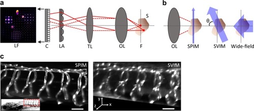
Selective volume illumination microscopy enhances LFM for the synchronous imaging of 3D samples.

a LFM is a simple extension of a conventional microscope, which produces a magnified image of the sample (S) from the native focal plane (F) to the image plane (IP) using an objective lens (OL,) and tube lens (TL). LFM places a micro-lens array (LA) at the IP, encoding 3D image information into a 2D light-field image (LF), which is captured by a planar detection camera. This permits LFM to synchronously capture information at z-positions above and below F; the 3D image of the sample is reconstructed from the LF image, based on knowledge of the optical transformation. b SVIM improves LFM by selectively illuminating the volume of interest within the sample. This decreases background and increases contrast when compared to wide-field illumination of the entire sample. SVIM was implemented through the use of light-sheet (SPIM) illumination that is scanned axially, so that the thin sheet of excitation is extended into a slab. In our work, the SVIM illumination axis was orthogonal to the detection axis (θ = 900), but the benefits of reduced background can be obtained by using illumination from a different angle, and/or by employing non-linear optical effects to selectively excite the volume of interest. c SPIM and SVIM 3D images of the trunk vasculature of 5 dpf zebrafish larva reveal the compromises between resolution and volumetric imaging time. SPIM offers higher resolution but requires the collection of 100 sequential images to cover the 100-µm-depth z-stack; SVIM captures the same 3D volume in a single snapshot, two-orders-of-magnitude faster, but with lower resolution. Transgenic animal, Tg(kdrl:GFP), had its vasculature fluorescently labeled with green fluorescent protein (GFP). Inset shows the approximate location of the imaged volume along the trunk of the zebrafish larva. Scale bars, 50 µm.

Higher contrast achieved by SVIM.

a SVIM images of the cranial vasculature improve in contrast as the depth (axial extent) of the illumination volume is decreased. Images are averaged-intensity z-projections of the same 40-µm thick sub-volume, centered at ~170 µm into the head of a 5 dpf zebrafish larva. The SVIM image quality progressively approaches the performance of SPIM as the axial extent of the illumination is reduced to 300 µm or 100 µm, far exceeding the image contrast obtained with wide-field LFM. Inset shows the approximate location of the imaged volume, in context of the zebrafish head. b Quantitative comparison of image contrast, defined as the normalized standard deviation of the pixel values (Methods section), comparing LFM, SPIM, and SVIM of different SVI extents from a. SVIM of smaller extents yielded increasingly better contrast, approaching the performance of SPIM. The contrast of SPIM showed the intrinsic contrast variation of the 3D sample, coupled with the expected contrast decay for increasing imaging depth. The local increase in contrast seen for the SVIM and LFM cases around z = 0 µm came from grid-like artifacts from the light-field reconstruction centered around the native focal plane, a known feature of LFM in general3,4. Scale bars, 100 µm.

SVIM enables fast, high-contrast, volumetric imaging of live biological systems.

ac Imaging the bacterial flow around the light organ of a juvenile squid. Raw light-field images recorded with conventional wide-field illumination yielded excessive background a, whereas SVIM, with a selectively illuminated volume of 100 µm, reduced this background and enhanced the contrast to allow localization of individual bacteria b. Inset shows squid with the light organ region highlighted by the dashed oval. c Quantitative flow trajectories tracked from the reconstructed SVIM data, color-coded for z-depth. Non-uniform 3D flow patterns were observed throughout the imaged volume. Images were collected at 20 volumes s−1, with a volume ~600 × 600 × 100 µm3 (depth). dg Imaging the motions of the beating heart wall and moving blood cells. A volume of ~250 × 150 × 150 µm3 (depth) in a live 5 dpf zebrafish larva was captured at 90 volumes s−1. Transgenes labeled the endocardium (rendered white) and blood cells (rendered red), Tg(kdrl:eGFP, gata1:dsRed). Inset in d highlights the position of the heart within the animal. The captured beating heart is shown in 3D-rendered views at two representative time points during the cardiac cycle: d the atrium was at its fullest expanded extent, followed by e when the blood had been pumped into the enlarged ventricle. Representative blood cell flow trajectories were manually tracked and quantified (color of the trajectories in d, e, g depicts blood cell speed; Methods section). f Maximum projection image along the x-axis of several representative flow trajectories highlights the substantial component of blood flow along the z-direction. To aid visualization, clipping planes in the yz plane were used to cut out the atrium and parts of the ventricle. Color-coding of the blood cell tracks in f is only for visual identification. g Perspective view of the blood cells demonstrates the achieved single-cell resolution, notably along the z-direction. Circular voids within several blood cells mark the cells whose trajectories were tracked and quantified. Scale bars, (ac) 100 µm, (dg) 50 µm.

Functional neuroimaging with SVIM.

Functional imaging of a 5 dpf larval zebrafish with pan-neuronal fluorescent calcium indicators, Tg(elavl3:H2b-GCaMP6s). Spontaneous brain activity, over a volume ~600 × 600 × 100 µm3 (depth), was recorded at 1 volume s−1, with SVIM, in either 1- or 2-photon excitation mode (1p-SVIM or 2p-SVIM, respectively), or conventional 1p wide-field LFM. Cellular-resolution representations of active neurons were found with standard methodology based on spot segmentation of the time-domain standard deviation of the 3D time-series data (Methods section). ac Images shown are depth color-coded xy- or xz-projections, of the time-domain standard deviation projection of the recorded brain activity over a time window of 100 s. Colored ellipsoids represent active neurons. Dashed box in the xy-projection image represent the region that produces the corresponding xz-projection image. Activity traces of segmented neurons are shown in df, revealing that the most number of neurons were found with 2p-SVIM (1104 cells), then with 1p-SVIM (796 cells), both of which were several-fold higher than with conventional wide-field LFM (263 cells). Scale bars, 100 µm.

Acknowledgments
This image is the copyrighted work of the attributed author or publisher, and ZFIN has permission only to display this image to its users. Additional permissions should be obtained from the applicable author or publisher of the image. Full text @ Commun Biol