FIGURE SUMMARY
Title

Midkine-a is required for cell cycle progression of Müller glia glia during neuronal regeneration in the vertebrate retina

Authors
Nagashima, M., D'Cruz, T.S., Danku, A.E., Hesse, D., Sifuentes, C., Raymond, P.A., Hitchcock, P.F.
Source
Full text @ J. Neurosci.

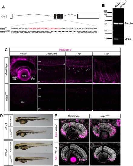
Midkine-a governs cell cycle kinetics during retinal development. A, Schematic representation of the midkine-a locus on chromosome 7. The gene consists of five exons. Red represents the gRNA target sequence located at exon 3. B, Using larvae at 48 h post fertilization (hpf), Western blot analysis for Midkine-a confirms lack of protein in the mdkami5001 mutant. C, Immunocytochemistry for Midkine-a in embryonic (48 hpf), adult, and regenerating retinas. In unlesioned retina, Midkine-a immunoreactivity is detected in horizontal cells (arrowhead) (Gramage et al., 2015). Photoreceptor cell death induces Midkine-a in reprogrammed Müller glia (arrows) at 1 dpl. Midkine-a distributes radial processes of Müller glia at 3 dpl. D, Lateral views of larvae at 48 and 72 hpf: AB-WT and mdkami5001 mutant. E, Proliferation assay with EdU labeling at 48 hpf. Compared with WT, there is an increased number of EdU-labeled cells in the retinas of mdkami5001 mutants. Retinal lamination and cellular differentiation are delayed, but not blocked in the mdkami5001 at 72 hpf. cmz, Ciliary marginal zone; onl, outer nuclear layer; inl, inner nuclear layer; gcl, ganglion cell layer. Scale bar, 40 μm. (see extended data, Table 1-1, and Table 1-2).

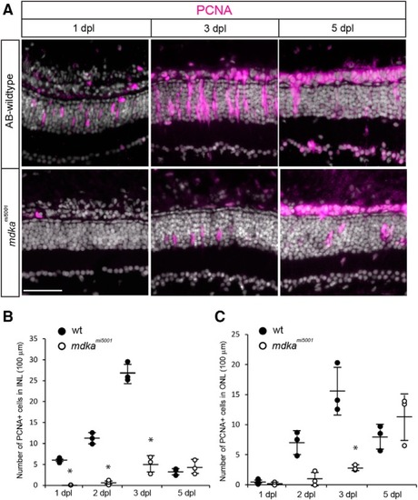
In the mdkami5001 mutant, Müller glia fail to proliferate in response to photoreceptor cell death. A, Immunocytochemistry for PCNA (magenta) in WT and mdkami5001 at 1 and 3 dpl. In WT, Müller glia become positive for PCNA at 1 dpl. At 3 dpl, PCNA+ progenitors form neurogenic clusters. Mutant retinas lack PCNA-labeled cells at 1 dpl. Very few, isolated cells are positive for PCNA in the mdkami5001 at 3 dpl. B, C, The number of PCNA+ cells in WT and mdkami5001 in the inner (B; 1 dpl: p = 0.0017; 2 dpl: p < 0.0001; 3 dpl: p < 0.0004, ANOVA with post hoc Tukey; F ratio = 116.1834) and outer (C; 3 dpl; p = 0.0001, ANOVA with post hoc Tukey; F ratio = 17.5589) nuclear layers. The mdkami5001 retinas have significantly less PCNA+ cells at 1, 2, and 3 dpl compared with WT retinas. A total of 3 sections were counted and averaged in each retina. A total of 3 retinas were analyzed. Scale bar, 30 μm. *p < 0.01.

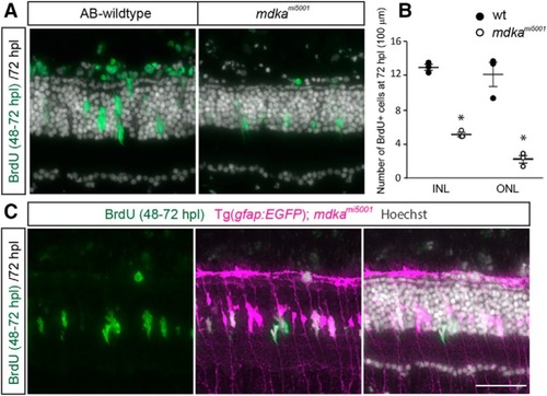
Some Müller glia in the mdkami5001 progress through the cell cycle. A, B, BrdU labeling between 48 and 72 hpl in WT and mdkami5001 mutant retinas. In WT, Müller glia-derived progenitors are labeled with BrdU. In mdkami5001 mutant at 72 hpl, fewer cells in the inner nuclear layer (INL) and outer nuclear layer (ONL) are labeled with BrdU compared with WT (INL: p < 0.0001; ONL: p = 0.0008, ANOVA with post hoc Tukey). C, BrdU labeling (green) in transgenic reporter line, Tg(gfap:eGFP)mi2002, in the mdkami5001 mutant background. Tg(gfap:eGFP)mi2002 is pseudocolored in magenta. Scale bar, 30 μm. *p < 0.01.

The mdkami5001 mutant retinas fail to regenerate cone photoreceptors. A, Immunocytochemistry for red/green cone photoreceptor marker, Zpr1. In WT retina, immature cone photoreceptors start to appear at 5 dpl, and regeneration largely completes by 14 dpl. In the mdkami5001 mutant, regenerating photoreceptors are absent at 5 dpl. At 7 dpl, very few cone photoreceptors appear. The number of cone photoreceptors is less at 14 dpl compared with WT. B, Immunocytochemistry for rod photoreceptor marker Zpr3 following lesion. In WT retina, regenerating rod photoreceptors appear by 7 dpl. In the mdkami5001 retinas, rod photoreceptors slowly regenerate by 28 dpl. C, Flat-mounted retinal preparation immunostained with ZO1 in unlesioned and 14 dpl. In unlesioned retina of both WT and mdkami5001, cone photoreceptors form a crystalline mosaic array in the planar apical surface of the retina (Livak and Schmittgen, 2001; Nagashima et al., 2017). Higher magnification of boxed region indicates the alignment of cones in the mosaic array (asterisks) with flattened cell boundaries (arrowheads). At 14 dpl in WT, cone photoreceptors regenerate (asterisks), although the crystalline mosaic array is not restored. In the mdkami5001 retina, cone profiles are instead replaced by irregularly shaped, expanded Müller glial apical processes (dotted line). D, Counts of ZO1-labeled cone photoreceptors at 14 and 28 dpl. Significantly fewer cones are regenerated in the mdkami5005 mutant (white) compared with WT (gray). n = 6. 14 dpl: p = 0.0051; 28 dpl: p = 0.0051, nonparametric Mann-Whitney-Wilcoxon. onl, Outer nuclear layer; inl, inner nuclear layer; gcl, ganglion cell layer. Scale bars: A, B, 30 μm; C, 10 μm. *p < 0.01.

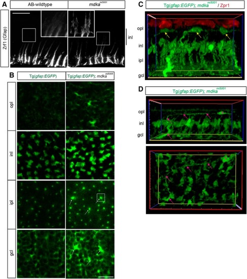
Following photoreceptor death, Müller glia in the mdkami5001 mutant undergo gliotic remodeling. A, Immunocytochemistry for Gfap in WT and mdkami5001 retinas at 28 dpl. In WT, the Gfap immunosignal is restricted to the inner third of radial processes. No obvious signal is detected at the inner nuclear layer. The mdkami5001 upregulates Gfap, and signals are seen at the cell body of Müller glia in the inner nuclear layer. B, Single optical planes from z-stack series of the Tg(gfap: EGFP) reporter flat-mount retinal preparation in the mdkami5001 background. In the ganglion cell and inner plexiform layers, some Müller glia show signs of hypertrophy, including increased levels of the EGFP transgene signal (arrows). C, Cross section view of 3D reconstructed image in the Tg(gfap:GFP); mdkami5001 (green) retina at 28 dpl, immunolabeled with Zpr1 (red) in a flat-mount preparation. Yellow arrows indicate displaced Müller glia somata in the outer plexiform layer. D, Cross section and flat-mounted views of the 3D reconstructed image. Displaced Müller glia (magenta asterisk) retain basal radial process (magenta arrows). opl, Outer plexiform layer; inl, inner nuclear layer; ipl, inner plexiform layer; gcl, ganglion cell layer. Scale bars: A, 30 μm; B, 20 μm.

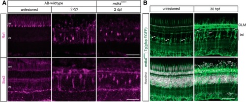
Müller glia in the mdkami5001 mutant dedifferentiate following photoreceptor death. A, Immunocytochemistry for regeneration-associated genes, Rx1 and Sox2, following photolytic lesion in WT and mdkami5001 retinas. Lesion induces Rx1 and Sox2 expression in Müller glia both in WT and mdkami5001 retinas at 2 dpl. B, The mdkami5001 retinas carrying the Müller glial reporter, Tg(gfap:eGFP)mi2002. Photoreceptor injury induces interkinetic nuclear migration of Müller glial nuclei in the mdkami5001 mutant. Arrows indicate cell bodies of Müller glia. The nuclei were stained with Hoechst (gray). OLM, Outer limiting membrane; inl, inner nuclear layer; onl, outer nuclear layer. Scale bars, 30 μm.

The mdkami5001 mutant upregulates “core” transcriptional regulators following photoreceptor death. A–C, qPCR for dedifferentiation markers, ascl1a, stat3, and lin28, at 30 and 36 hpl. Both WT and mdkami5001 upregulate ascl1a (A; WT: 30 hpl, p < 0.0001, 36 hpl, p < 0.0001; mdkami5001: 30 hpl, p = 0.0004, 36 hpl, p = 0.006, p = 0.0066 relative to WT; F-ratio = 37.5606), lin28 (B; WT: 30 hpl, p < 0.0001, 36 hpl, p = 0.0095; mdkami5001: 30 hpl, p < 0.0001, 36 hpl, p = 0.0004; F-ratio = 32.9337), and stat3 (C; WT: 30 hpl, p = 0.0048, 36 hpl, p = 0.0016; mdkami5001: 30 hpl, p = 0.0004, 36 hpl, p = 0.0016), following lesion. ANOVA with post hoc Tukey, relative to unlesioned. D, E, Western blot for Stat3 in WT and mdkami5001 retinas at 1 dpl. WT: p = 0.0002, mdkami5001: p = 0.0004, ANOVA with post hoc Tukey; F-ratio = 40.8763. F, Immunocytochemistry for phosphorylated Stat3 (pStat3) in the WT and mdkami5001 mutant. In unlesioned retina, immunosignal for pStat3 is not detected in the inner nuclear layer. Following photoreceptor lesion, Müller glia in the inner nuclear layer upregulate pStat3 in WT, whereas mdkami5001 mutants have reduced phosphorylation of Stat3. Scale bar, 30 μm. inl, Inner nuclear layer; ipl, inner plexiform layer. *p < 0.01.

Müller glia in the mdkami5001 mutant arrest in the G1 phase of the cell cycle. A–C, qPCR assay for cell cycle regulator cyclins, ccnd1, ccne1, and ccna2 following photolytic lesion. Both WT and mdkami5001 upregulate G1 cyclins, ccnd1 (A; WT: 30 hpl, p = 0.0107, 36 hpl, p = 0.0013; mdkami5001: 30 hpl, p = 0.0048, 36 hpl, p = 0.0213; F-ratio = 12.0576) and ccne1 (B; WT: 30 hpl, p < 0.0001, 36 hpl, p < 0.0001; mdkami5001: 30 hpl, p = 0.0012, 36 hpl, p = 0.0001; F-ratio = 36.5808), following lesion (A,B). The mdkami5001 mutant fails to upregulate S phase cyclin, ccna2 (C; WT: 36 hpl, p = 0.0011; F- ratio = 12.5433). D, S phase assay with BrdU labeling (green) between 24 and 30 hpl. Müller glia in the mdkami5001 mutants did not incorporate BrdU following lesion. E–H, qPCR of additional cell cycle regulators. Expression of cyclin-dependent kinases, cdk4 (E; WT: 36 hpl, p < 0.0001; F-ratio = 15.8783) and cdk6 (F; WT: 36 hpl, p = 0.0087; F- ratio = 8.3300), is dysregulated in the mdkami5001 retinas (E,F). The WT transiently upregulates id2a at 30 hpl (p = 0.0003; F- ratio = 7.2578), whereas expression levels did not change in the mdkami5001 (G). Expression of the cell cycle inhibitor, p130, decreases in the WT after lesion, whereas mdkami5001 maintains steady levels of expression (H; p = 0.002, relative to WT; F- ratio = 6.3823). ANOVA with post hoc Tukey, relative to unlesioned. Scale bar: D, 30 μm. onl, Outer nuclear layer; inl, inner nuclear layer; gcl, ganglion cell layer. *p < 0.03, #p < 0.01.

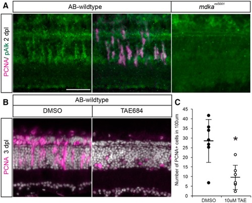
Activation of the ALK receptor is required for Müller glial to proliferate. A, Double immunostaining for PCNA (magenta) and pAlk (green). PCNA+ cells express pAlk in WT retina at 2 dpl, whereas pAlk immunostaining was not detected in mdkami5001 retina. Scale bar, 30 μm. B, Pharmacological inhibition of Alk using TAE684 suppresses proliferation in the WT retinas after a lesion. C, Counts of PCNA+ proliferative cells in DMSO- or TAE684-treated WT retinas at 3 dpl. n = 7. *p = 0.0011 (nonparametric Mann-Whitney-Wilcoxon).

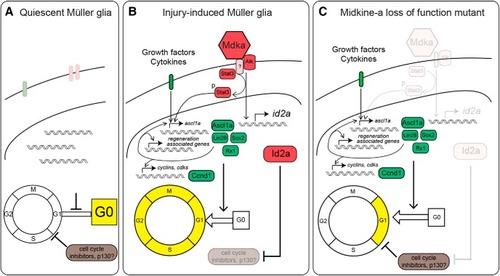
Model of Midkine-a-mediated cell cycle regulation in Müller glia. A, In quiescent Müller glia, cell cycle inhibitors keep cells in G0 phase. B, Injury induces cytokines and growth factors to upregulate regeneration-associated reprogramming genes for dedifferentiation and cell cycle reentry. Midkine-a-Alk signaling participates in the induction of the regeneration-associated gene, ascl1a, via phosphorylation of Stat3. Midkine-a signaling also induces expression of the cell cycle regulator, id2a, that inhibits cell cycle inhibitors, such as p130. C, In the absence of Midkine-a, Müller glia fail to suppress cell cycle inhibition, resulting in compromised progression of the cell cycle.

Acknowledgments
This image is the copyrighted work of the attributed author or publisher, and ZFIN has permission only to display this image to its users. Additional permissions should be obtained from the applicable author or publisher of the image. Full text @ J. Neurosci.