FIGURE SUMMARY
Title

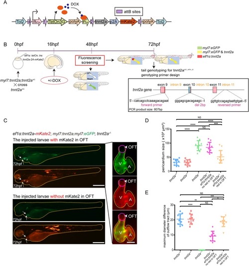
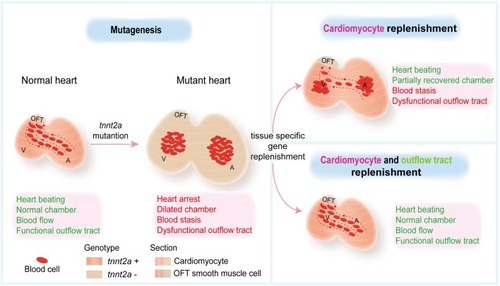
Combinatorial genetic replenishments in myocardial and outflow tract tissues restore heart function in tnnt2 mutant zebrafish

Authors
Liu, L., Fei, F., Zhang, R., Wu, F., Yang, Q., Wang, F., Sun, S., Zhao, H., Li, Q., Wang, L., Wang, Y., Gui, Y., Wang, X.
Source
Full text @ Biol. Open

Generation of tnnt2a mutant zebrafish andphenotype analyses. (A) Graphical representation of the target site of tnnt2a for CRISPR/Cas9 gRNA and altered amino acid sequence of Tnnt2a of mutant zebrafish. (B) Genotype sequencing results of tnnt2a+/+ and tnnt2a−/− zebrafish. (C) Representative images of atrial (A) and ventricular (V) enlargement, and pericardial effusion in 72 hpf tnnt2a−/− zebrafish. Scale bars: 150 µm. (D–G) Statistics for the heart rate, atrial and ventricular diameter, and pericardium size in wild-type and tnnt2a−/− zebrafish; ****P<0.0001; n=15 per group. Statistical differences were evaluated using two-tailed unpaired t-tests (D–F) or unpaired t-test with Welch's correction (G). (H,I) Fluorescent images and pseudo-colours of electrocardiac rhythm in Tg(bactin2:GCamp6s; tnnt2a+/+) and Tg(bactin2:GCamp6s; tnnt2a−/−) zebrafish; arrows represent increased Ca2+ flux in cardiomyocytes. The direction of Ca2+ flux was from venous to arterial end both in tnnt2a+/+ and tnnt2a−/−. Scale bars: 100 µm.

Myocardial-specific and DOX-inducible tnnt2a expression fail to rescue heart function in tnnt2a mutant zebrafish. (A) Structure of the tol2-flanked transgenic vector. DOX, doxytetracycline. (B) Lateral views of a 72 hpf transgenic zebrafish after DOX induction at 48 hpf in green and red fluorescence. Scale bar: 300 µm. (C) Real-time RT-PCR of wild-type form of tnnt2a cDNA in wild-type, tnnt2a−/−, Tg(myl7:tetOn; tre:tnnt2a-p2a-mKate2; myl7:eGFP) [Tg(myl7:tnnt2a)] following addition of DOX at 16 hpf, and Tg(myl7:tetOn; tre:tnnt2a-p2A-mKate2; myl7:eGFP; tnnt2a−/−) [Tg(myl7:tnnt2a); tnnt2a−/−] following addition of DOX at 16 or 48 hpf. **P<0.01, ***P<0.001, ****P<0.0001, NS, not significant; n=30 per group. Statistical differences were evaluated using two-way ANOVA. (D) Representative images of hearts of four groups from ventral view. Yellow dotted line, pericardium; white line, heart outline; black box, area of the fluorescent images; V, ventricle; A, atrium. Scale bar: (bright field) 50 µm, (fluorescent) 100 µm. DOX was added at 16 hpf. (E–H) Statistics for heart rate, atrial and ventricular end-diastolic diameter, and pericardium size in four groups; **P<0.01, ****P<0.0001, NS, not significant; n=15 per group. Statistical differences were evaluated using Kruskal–Wallis test (E) or one-way ANOVA (F–H).

Gene expression in OFT is deregulated in tnnt2a mutant zebrafish with myocardial tnnt2a supplementation. (A) Workflow of sample collections for whole-fish RNA-seq of four groups: tnnt2+/−, tnnt2a−/− Tg(myl7:tnnt2a); tnnt2a−/− and Tg(myl7:tnnt2a); tnnt2a+/−. (B) Volcano plot of differentially expressed genes in tnnt2a+/− versus Tg(myl7:tnnt2a); tnnt2a−/−. (C,D) Sarcomere- and OFT-relevant gene expression in dissected heart of tnnt2a+/+, tnnt2a−/−, and Tg(myl7:tnnt2a); tnnt2a−/− after DOX induction at 16 hpf. *P<0.05, ***P<0.001, ****P<0.0001, NS, not significant; n=30 per group. Statistical differences were evaluated using two-way ANOVA. Red asterisks, sarcomere genes in Tg(myl7:tnnt2a); tnnt2a−/− can almost return to wild-type level after DOX induction; red hashes, OFT-relevant genes in Tg(myl7:tnnt2a); tnnt2a−/− are more deviated from wild-type level after DOX induction compared with tnnt2a−/−.

Mechanical behaviours of OFT are deregulated in tnnt2a mutant zebrafish with myocardial tnnt2a supplementation. (A) Representative images of the OFT morphologies at systole and diastole stages of a cardiac cycle. V, ventricle; A, atrium; yellow dotted line, heart outline; white line, OFT diameter; purple dotted line, OFT outline. Scale bar: 100 µm. (B) Statistics of maximum diameter difference of OFT in tnnt2a+/+, tnnt2a−/− and Tg(myl7:tnnt2a); tnnt2a−/−. ****P<0.0001; n=30 per group. Statistical differences were evaluated using Kruskal–Wallis test. (C) Images of the stacked blood cells in the heart chambers of tnnt2a−/− and Tg(myl7:tnnt2a); tnnt2a−/− zebrafish at 72 hpf. Arrows show the stacked red blood cells. Scale bars: 100 µm.

Identification of the TNNT2-positive, Myl7-negative smooth muscle cells in the OFT. (A) Images of in situ hybridization with tnnt2a anti-sense probe. Yellow dotted line, outflow tract outline; green dotted line, cell outline. (B) Immunofluorescence of Tnnt2, Myl7, and α-SMA staining in wild-type zebrafish hearts at 5 dpf. Nuclei is stained by Hoechst 33342. Yellow circle shows OFT area. (C) Myl7 zebrafish heart stained by phalloidin (F-actin) at 5 dpf. Nuclei is stained by Hoechst 33342. Scale bar: 150 µm. (D,E) Images of a single plane of Tnnt2, Myl7, α-SMA and phalloidin immunofluorescence staining. V, ventricle; A, atrium. Scale bars: (A–C) 150 µm, (D,E) 50 µm.

Replenishments of tnnt2a in both myocardial cells and OFT significantly recover the heart function of tnnt2a mutant zebrafish. (A) Structure of the transgenic vector for rescue injection. (B) Schematic workflow of the double-rescue experiments and phenotyping/genotyping. (C) Left: representative images of Tg(myl7:tnnt2a); tnnt2a−/− zebrafish injected by eflα:tetOn; tre:tnnt2a-p2A-mKate2 [eflα:tnnt2a] with and without red fluorescence in OFT. Right: representative images of Tg(myl7:tnnt2a); tnnt2a−/− with additional eflα:tnnt2a in OFT. Yellow dotted lines, whole larvae or heart outlines; arrows and purple circle, mKate2-positive and eGFP-negative regions; V, ventricle; A, atrium; arrows and arrowheads, outflow tract. Scale bars: (lateral view) 200 µm, (ventral view) 150 µm. (D,E) Statistics of the maximum diameter difference of the OFT and pericardium size in tnnt2a+/+, tnnt2a+/−, tnnt2a−/−, Tg(myl7:tnnt2a); tnnt2a−/− injected by eflα:tetOn; tre:tnnt2a-p2A-mKate2 [eflα:tnnt2a] with and without red fluorescence in OFT. *P<0.05, **P<0.01, ***P<0.001, ****P<0.0001, NS, not significant; n=30 per group. Statistical differences were evaluated using one-way ANOVA (D) and Kruskal–Wallis test (E).

Graphical abstract. Myocardial-specific genetic replenishments are insufficient to rescue tnnt2 mutant heart function, and combinatorial genetic replenishments in myocardial and OFT tissues restore heart function in tnnt2 mutant zebrafish.

Acknowledgments
This image is the copyrighted work of the attributed author or publisher, and ZFIN has permission only to display this image to its users. Additional permissions should be obtained from the applicable author or publisher of the image. Full text @ Biol. Open