
- Title
-
A robust human norovirus replication model in zebrafish larvae
- Authors
- Van Dycke, J., Ny, A., Conceição-Neto, N., Maes, J., Hosmillo, M., Cuvry, A., Goodfellow, I., Nogueira, T.C., Verbeken, E., Matthijnssens, J., de Witte, P., Neyts, J., Rocha-Pereira, J.
- Source
- Full text @ PLoS Pathog.

( |
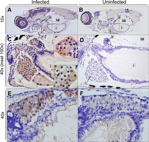
Immunohistochemistry of HuNoV GII.P7-GII.6-infected zebrafish larvae ( |
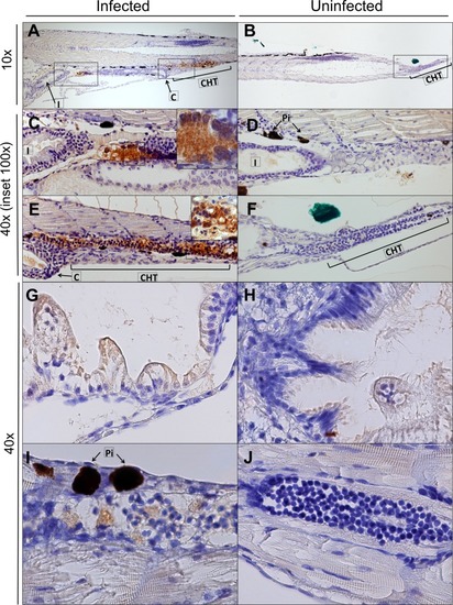
Immunohistochemistry of HuNoV GII.P7-GII.6-infected zebrafish larvae ( |