- Title
-
Mating Suppresses Alarm Response in Zebrafish
- Authors
- Diaz-Verdugo, C., Sun, G.J., Fawcett, C.H., Zhu, P., Fishman, M.C.
- Source
- Full text @ Curr. Biol.
|
Independent, Non-Interacting Mating-Water and Skin-Extract Activation Patterns in the Olfactory Bulb (A) Mean response to olfactory stimulation in two optical sections at 0 μm (top) and 80 μm (bottom) depth of the left olfactory bulb (OB) of a 32-day-old juvenile tuba1a:GCaMP6s zebrafish, when exposed to skin extract and the mixture of skin extract and mating water. Ellipses outline representative glomeruli: dorsal (dG) and lateral (lG). Right panel: mean ΔF/F signal traces of odor responses of the glomerulus region of interest annotated in the section. (B) Mean response to olfactory stimulation in two optical sections at 128 μm (top) and 192 μm (bottom) depth of the left OB of the same fish as in (A) and when exposed to mating water and the mixture of skin extract and mating water. Ellipses outline representative glomeruli: medio-anterior (maG) and ventromedial glomerular (vmG) clusters. Right panel: mean ΔF/F signal traces of odor responses of the glomerulus region of interest annotated in the section. (C) Mean ΔF/F signal traces of odor responses within each OB glomerulus region of interest annotated in (A) (n = 7 fish). Shaded regions denote the standard error of the mean. (D) Mean ΔF/F signal traces of odor responses within each OB glomerulus region of interest annotated in B (n = 7 fish). Shaded regions denote the standard error of the mean. See also Figure S2 and Video S3. lG, lateral glomerular; dG, dorsal glomerular; maG, medio-anterior glomerular; vmG, ventro-medial glomerular; A, anterior; P, posterior; M, medial; L, lateral (L). Scale bar, 50 μm. |
|
Skin Extract and Mating Water Elicit Responses in Both Specific and Overlapping Telencephalic Areas (A) Anatomical reference (upper panel schematic) of 2-photon imaged brain regions (lower panel images) in 34-day-old juvenile tuba1a:GCaMP6s zebrafish. Each colored dot represents an roi activated in response either to one stimulus specifically (magenta, mating water; green, skin extract), to both stimuli (white), or to the mating-water and skin-extract stimulus mixture (cyan). Red-dotted regions contain skin-extract-selective rois that were suppressed when given the stimulus mixture (Dp and posterior-lateral Dm). Yellow-dotted region delineates Vs, which contain mating-water-selective rois, whose activity persisted when given the stimulus mixture. (B) Left panel: mean ΔF/F signal of mating-water-selective rois in the telencephalon (n = 4,104 rois, n = 6 fish). Right panel: mean ΔF/F signal of skin-extract-selective rois in the telencephalon (n = 4,109 rois, n = 6 fish). (C) ΔF/F signal of skin-extract- (top rows) or mating-water- (bottom rows) selective rois in a 20-s window post-delivery of mating water, skin extract, or mixture stimulus (n = 8,213 rois, n = 6 fish). See also Figure S3. Tel, Telencephalon; OB, olfactory bulb; Hb, habenula; OT, optic tectum; DI, lateral zone of the dorsal telencephalic area; Dm, medial zone of the dorsal telencephalic area; Dp, posterior part of the dorsal telencephalic area; Vs, supracommisural nucleus of the ventral telencephalic area; and Vp, postcommisural nucleus of the ventral telencephalic area. Scale bar, 50 μm. |
|
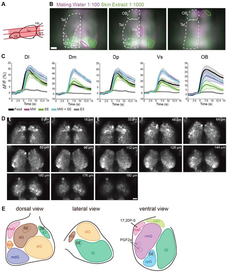
Widefield calcium imaging of olfactory bulb and telencephalon reveals brain area responding to mating water and skin extract. Olfactory bulb anatomy. Related to Figure 2. (A) Schematic representation of the forebrain of a juvenile zebrafish brain showing the anatomical position of the three optical sections imaged. (B) Mean ΔF/F signal in response to mating water (magenta) and skin extract (green; n = 5 fish). From left to right, in order from dorsal to ventral: three imaged optical sections. Solid magenta and green lines denote prominent areas that responded primarily to mating water and skin extract, respectively. (C) Mean ΔF/F signal time course in five representative areas (Dl, Dm, Dp, Vs and OB) for each odor delivered. Shaded regions represent the standard error of the mean. (D) Anatomical reference of 2-photon imaged olfactory bulb in 34 day-old juvenile tuba1a:GCaMP6s zebrafish. Number at the top right corner indicates the imaging depth. E) Schematic representation of the odor map and glomerular clusters of the olfactory bulb. Figure adapted from Yoshihara, 2014 [S1]. Abbreviations: Telencephalon (Tel), olfactory bulb (OB), lateral zone of the dorsal telencephalic area (Dl), medial zone of the dorsal telencephalic area (Dm), posterior part of the dorsal telencephalic area (Dp), and supracommisural nucleus of the ventral telencephalic area (Vs), dorsal glomerular (dG), dorso-lateral glomerular (glG), lateral (lG), medio-anterior (maG), medioposterior (mpG), medio-dorsal (mdG), ventro-anterior (vaG), ventro-medial (vmG), ventroposterior (vpG) glomerular cluster. Scale = 50 μm. |
|
Raw data from 2-photon calcium imaging of telencephalon. Related to Figure 3. (A) Odor-evoked activity map (mean F/F signal during 5 s post-stimulus delivery) of the telencephalic section in B to different stimuli: food, control (E3), mating water, skin extract and mixture of mating water and skin extract. (B) Left: anatomical section of 2-photon imaged telencephalon in 34 day-old juvenile tuba1a:GCaMP6s zebrafish with two sample rois (1, 2) highlighted. Right: mean ΔF/F signal of the two sample rois to each stimulus. Scale = 50μm. |


