- Title
-
Neuropilin 1 mediates epicardial activation and revascularization in the regenerating zebrafish heart
- Authors
- Lowe, V., Wisniewski, L., Sayers, J., Evans, I., Frankel, P., Mercader-Huber, N., Zachary, I.C., Pellet-Many, C.
- Source
- Full text @ Development
|
Nrps are upregulated during zebrafish heart regeneration. (A) Absolute qPCR analysis of nrp family genes at 1, 3, 7, 14, 30 and 60 days following cryoinjury or sham surgery. Basal expression was evaluated in uninjured hearts of age-matched wild-type fish. Bars represent normalized copy number per reaction. Data are mean±s.e.m. **P<0.01, ***P<0.005, ****P<0.001 (one-way ANOVA with Sidak's post hoc test for multiple comparisons of n=4 or 5 with each n being a pool of five ventricles). (B) Adult zebrafish ventricle lysates 1, 3, 7, 14 and 30 days following surgery, immunoblotted for Nrp1 and Gapdh (left); western blot quantification of Nrp1 protein in sham and cryoinjured ventricles 1, 3, 7, 14 and 30 days following surgery (right) (n=4 or 5, with each nbeing a pool of three ventricles). (C) In situ hybridization with digoxigenin-labeled antisense riboprobes were used to detect nrp family isoforms in sham-operated and cryoinjured adult zebrafish hearts 1, 3 and 14 dpci (n≥3). Arrowheads indicate gene expression within the epicardium. CI, cryoinjured; epi, epicardium; IA, injured area. Scale bars: 250 µm. |

ZFIN is incorporating published figure images and captions as part of an ongoing project. Figures from some publications have not yet been curated, or are not available for display because of copyright restrictions. EXPRESSION / LABELING:
|
|
Characterization of nrp1asa1485 mutant fish. (A) Structure of Nrp1a in wild-type (nrp1a+/+) (left) and nrp1asa1485 mutant fish (right). The point mutation results in the generation of a premature stop codon at aa 206, resulting in a truncated Nrp1a fragment. Blue diamonds indicate CUB (a1 and a2) domains, orange circles indicate FA58C (b1 and b2) domains, the green square indicates the MAM domain and the brown square indicates the C-terminal domain. (B) Sequencing chromatograms of wild-type fish, heterozygous nrp1asa1485/+ and homozygous nrp1asa1485/sa1485 mutant fish. An early stop codon (nonsense mutation) TAA, replaces the wild-type TAC codon at aa 206. The genotypes of 14 zebrafish embryos 48 h post fertilization (hpf) were compared against the expected Mendelian ratio after heterozygous fish incross. (C) Absolute RT-qPCR of wild-type (WT; black circles, white bar) or nrp1asa1485 homozygous mutant (black squares, gray bar) uninjured adult zebrafish hearts under basal conditions. nrp1aexpression was significantly decreased in nrp1asa1485 samples, suggesting nonsense-mediating decay. ****P<0.0001 (two-tailed t-test; n=7 with each n being a pool of three ventricles). Data are means of normalized copy numbers per reaction±s.e.m. (D) Nrp1a antisense (AS) in situhybridization of wild-type (upper row) or nrp1asa1485 homozygous mutant (lower row) embryos 24 hpf. Nrp1a expression was clearly decreased in nrp1asa1485 samples. Scale bars: 50 μm. (E) Western blot of wild-type (WT) or nrp1asa1485 homozygous mutant uninjured adult zebrafish ventricle lysates (top). Lysates were immunoblotted with an antibody targeting the Nrp1 cytoplasmic domain and Gapdh. Note the absence of C terminus detection of Nrp1a in the nrp1asa1485 samples. Western blot quantification of Nrp1a (plain bars, circles for wild type, squares for nrp1asa1485) and Nrp1b (striated bars, upward triangles for wild type, downward triangles for nrp1asa1485) normalized to Gapdh (one-way ANOVA with Sidak's post hoc test for multiple comparisons of n=4), confirming the significant reduction in Nrp1a expression (****P<0.0001; plain white bar for wild type versus plain gray bar for nrp1asa1485), whereas Nrp1b was not significantly different between wild type (white striated bar) and nrp1asa1485 (gray striated bar) (P=0.219) (bottom). |
|
Cardiac regeneration is delayed in nrp1asa1485 mutants following cryoinjury. (A) Heart sections from wild-type (top) and nrp1asa1485 mutant (bottom) fish obtained at 1, 3, 7, 14, 30 and 60 dpci and stained with AFOG to identify the injured region. Dashed lines indicate the interface between cryoinjured and healthy tissue. (B) Cryoinjured areas were measured and represented as mean percentage of total ventricle area±s.e.m. P<0.05 (two-way ANOVA with Sidak's post hoc test for multiple comparisons of n=4-8). (C) A closed wound is one in which compact myocardium recovers following cryoinjury and encases the scar tissue; by contrast, a scar exposed to the surface is defined as an open wound. Wound closure was examined in wild-type (WT) and nrp1asa1485 mutant hearts at 30 and 60 dpci and open versus closed wounds were expressed as a percentage of the total number of hearts (n=4-8). (D,E) AFOG staining of wild-type and nrp1asa1485 mutant hearts at 3 dpci (D) used to evaluate epicardial thickness and injury boundaries to calculate the epicardial area normalized to the length of the injury boundary (continuous line), quantified in E. Dashed line represents outer boundary of epicardial area. *P<0.05 (two-tailed t-test of n=7 for wild-type and n=10 for nrp1asa1485 hearts). A, atrium; ba, bulbus arteriosus; V, ventricle. PHENOTYPE:
|
|
Neovascularization of the cryoinjured area is impaired in nrp1asa1485 mutants. (A,B) Blood vessels in either wild-type (A) or nrp1asa1485 (B) tg(fli1a:EGFP)y1 zebrafish at 1 (upper row) and 3 (lower row) dpci were identified in heart sections using GFP immunofluorescence in vascular structures. Heart sections were also counterstained with DAPI. Smaller images (left) represent DAPI staining (blue) and GFP staining (vessels, green) only; larger images (right) are the merged images. The white dashed line delineates the border of the area of injury. White arrowheads indicate blood vessels. (C,D) GFP-positive vessels were quantified at 1 dpci (C; **P<0.01, two-tailed t-test of n=3 and 4) and 3 dpci (D; *P<0.05, two-tailed t-test of n=4) for wild-type (white bars) versus nrp1asa1485 (gray bars) hearts. Individual data points (circles for wild type and squares for nrp1asa1485) represent individual hearts, each averaged from vessel counts in three to four different sections covering the injury site. |
|
Epicardial activation is decreased in nrp1asa1485 hearts following cryoinjury. (A-D) Wild-type and nrp1asa1485 mutant cryoinjured fish on the tg(wt1b:EGFP)li1 background were analyzed for epicardial activation at 3 dpci by identification of wt1b:EGFP-positive cells (A,C), and were also stained with DAPI (blue; A,C) and anti-PCNA antibodies (red; C), as indicated. In A, smaller images represent DAPI staining (blue) and GFP staining (activated epicardium, green) only; larger images are the merged images. In C, boxed area in left panel is enlarged in the middle. Smaller images (right) represent PCNA staining (red) and GFP staining (activated epicardium, green) only; larger images in the middle are the merged images. (B) Quantification of percentages of wt1b:EGFP-positive cells adjacent to the area of cryoinjury (indicated by the dashed line in A,C) in wild-type (white bar) and nrp1asa1485 (gray bar) mutant fish. **P<0.01 (two-tailed t-test of n=5). (D) Quantification of percentages of cells positive for wt1b:EGFP and PCNA in the area of cryoinjury. P>0.05 [two-tailed t-test of n=6 and 5 for wild-type (white bar) and nrp1asa1485 (gray bar), respectively]. Individual data points (circles for wild type and squares for nrp1asa1485) represent percentages in individual hearts, each averaged from counts in two to four different sections covering the injury borders. epi, epicardium; IA, injury area; ns, not significant. White-dotted line delineates injury–epicardial border. |

ZFIN is incorporating published figure images and captions as part of an ongoing project. Figures from some publications have not yet been curated, or are not available for display because of copyright restrictions. EXPRESSION / LABELING:
|
|
Epicardial cryoinjury-induced expansion and activation are impaired in nrp1asa1485 mutants. (A-D) The apices of wild-type and nrp1asa1485 zebrafish ventricles were collected 5 days post sham surgery or cryoinjury and cultured on fibrin gels for 7 days. (A) Epicardial cells migrated into the fibrin gels (dotted black lines). (B) Epicardial outgrowths were measured for each condition (sh, sham-operated and CI, cryoinjured hearts) after 7 days culture. Data are mean outgrowth area (mm2)±s.e.m. ***P<0.001 (one-way ANOVA with Sidak's post hoc test for multiple comparisons of n>9). (C) Epicardial explant recovered from wild-type and nrp1asa1485 tg(wt1b:EGFP)li1cryoinjured fish at 5 dpci were left to grow on fibrin gels for 7 days and stained for GFP. GFP fluorescence was observed at 10× (left column) and 40× magnification at the center and the periphery of the explants (middle and right columns, respectively). (D) Cell size (left) and ploidy (right) were quantified both at the center (top row) and at the edge (bottom row) of the explant. Data are expressed as percentage of cells per field of view ±s.e.m. Each n represents an average of 3 fields of view per explant (two-tailed t-test of n≥5); *P<0.05. ns, not significant. Scale bars: 1 mm in A, 20 µm in C (right); 100 µm in C (left). |

ZFIN is incorporating published figure images and captions as part of an ongoing project. Figures from some publications have not yet been curated, or are not available for display because of copyright restrictions. |
|
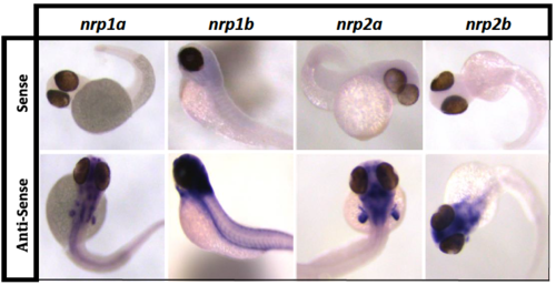
nrp riboprobes validation. In situ hybridization of TraNac transgenic zebrafish embryos 48 hours post fertilization (hpf) with nrp sense riboprobes (upper row) and nrp anti-sense riboprobes (lower row). Anti-sense riboprobes differential staining patterns were compared to previous reports (43) to confirm specific nrp isoform detection. All neuropilin isoforms are observed in the brain with additional differential expression patterns observed between different isoforms. Nrp1a is observed in the fin buds and otic vesicles, nrp1b is expressed in the dorsal aorta and intersegmental vessels, nrp2a is observed in the hind brain and fin buds, whereas nrp2b is largely restricted to the brain and hind brain, n ≥ 8. EXPRESSION / LABELING:
|
|
nrp1asa1485 mutant fish characterization (A) Representative picture of Wild-Type (upper panel) and nrp1asa1485 mutant zebrafish (lower panel), scale bar 1 cm. The body length (B), and heart size (C) of age matched Wild-Type (black dots) and nrp1asa1485 mutant (black squares) zebrafish were measured and compared (two-tailed t-test of n ≥ 5, p>0.05). (D) Scatter graph representing the values of body length to heart size ratio. Values are displayed as individual measurements of fish indicated as black dots (Wild-Type) or squares (nrp1asa1485). |

ZFIN is incorporating published figure images and captions as part of an ongoing project. Figures from some publications have not yet been curated, or are not available for display because of copyright restrictions. PHENOTYPE:
|


