
- Title
-
Glucocorticoids inhibit macrophage differentiation towards a pro-inflammatory phenotype upon wounding without affecting their migration
- Authors
- Xie, Y., Tolmeijer, S., Oskam, J.M., Tonkens, T., Meijer, A.H., Schaaf, M.J.M.
- Source
- Full text @ Dis. Model. Mech.

ZFIN is incorporating published figure images and captions as part of an ongoing project. Figures from some publications have not yet been curated, or are not available for display because of copyright restrictions. PHENOTYPE:
|
|
ZFIN is incorporating published figure images and captions as part of an ongoing project. Figures from some publications have not yet been curated, or are not available for display because of copyright restrictions. EXPRESSION / LABELING:
PHENOTYPE:
|
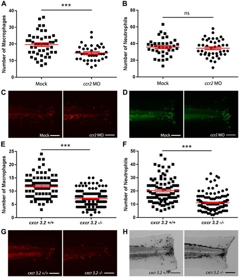
Effect of ccr2 morpholino knockdown or cxcr3.2 mutation on macrophage and neutrophil recruitment upon tail amputation in larvae. (A-B) The number of macrophages (A) and neutrophils (B) recruited to the wounded area at 4 hpa in 3dpf Tg(mpx:GFP/mpeg1:mCherry-F) larvae. In ccr2 morpholino-injected larvae, a significantly reduced number of macrophages were recruited compared to the number in mock (vehicle)-injected larvae. No significant difference was observed for the number of recruited neutrophils. (C-D) Representative images of the macrophages (fluorescently labeled by mCherry) (C) and the neutrophils (fluorescently labeled by GFP) (D) of mock-injected and ccr2 morpholino-injected larvae at 4 hpa. (E-F) The number of macrophages (E) and neutrophils (F) that recruited to the wounded area at 4 hpa in 3 dpf Tg(mpeg1:mCherry-F) larvae. A significantly reduced number of macrophages and neutrophils were recruited in cxcr3.2 mutant larvae compared to the number in wt controls. (G-H) Representative images of the macrophages (fluorescently labeled by mCherry) (G) and the neutrophils (stained using MPX assay) (H) of wt and cxcr3.2 mutant larvae at 4 hpa. Data are mean±s.e.m. (indicated in red), pooled from three independent experiments. ***P<0.001 (determined using the two-tailed t-test). ns, non-significant. Scale bars: 100 μm.
EXPRESSION / LABELING:
PHENOTYPE:
|
|
ZFIN is incorporating published figure images and captions as part of an ongoing project. Figures from some publications have not yet been curated, or are not available for display because of copyright restrictions. EXPRESSION / LABELING:
PHENOTYPE:
|
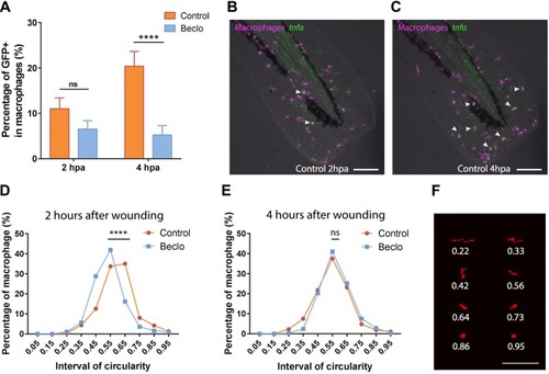
Effect of beclomethasone on the phenotype of macrophages. (A) In Tg(mpeg1:mCherry-F/tnfa:eGFP-F) reporter larvae, the number of GFP-positive macrophages (as a percentage of the total number of macrophages) recruited to the wounded area was quantified at 2 and 4 hpa in 5 dpf larvae. In the beclomethasone-treated group (Beclo), at 4 hpa, a significantly reduced percentage of the recruited macrophages was GFP-positive compared to the vehicle-treated group (Control). Data are mean±s.e.m. ****P<0.0001 (determined using ANOVA with a Fisher's LSD post hoc). (B-C) Representative images of macrophages (fluorescently labeled by mCherry) and GFP-positive macrophages (fluorescently labeled by both mCherry and GFP) in the control group at 2 hpa (B) and 4 hpa (C). Arrowheads indicate macrophages displaying the GFP signal, which is a measure for activation of the tnfa promoter. (D-E) The distribution of circularity of macrophages recruited to the wounded area at 2 h after wounding (hpa) (D) and at 4 h after wounding (E) in 3 dpf Tg(mpeg1:mCherry-F) larvae. At 2 hpa, a significant difference of distribution pattern was observed between the two groups, with the beclomethasone-treated group (Beclo) shifted towards lower circularity. At 4 hpa, no significant difference was observed. ****P<0.0001 (determined using the Kolmogorov–Smirnov test). (F) Representative images of macrophages analyzed in D and E and their corresponding circularity. ns, non-significant. Scale bars: 100 μm.
EXPRESSION / LABELING:
PHENOTYPE:
|