
- Title
-
Modeling Cancer with Flies and Fish
- Authors
- Cagan, R.L., Zon, L.I., White, R.M.
- Source
- Full text @ Dev. Cell

Examples of Tumors in Flies and Fish (A) RASG12V; PTEN−/− double mutant clones formed large glial-derived tumors in the fly brain. Red, repo (all glia); green, GFP (transformed glia). Insert shows the abnormally high density of glial-cell nuclei. From Read et al., 2009. (B) High magnification view of a multi-target (involving four mutations) cancer model achieved by overexpressing RASG12V and using RNAi-mediated knockdown of PTEN, APC, and P53. Green, transformed hindgut cells; red, muscle cells; and blue, trachea. See Bangi et al., 2016. (C) A transgenic zebrafish melanoma model. Expression of BRAFV600E under the melanocyte-specific mitfa promoter resulted in stripe disruption and nevi. When crossed with p53-mutant fish, the resultant mitfa-BRAFV600E;p53−/− fish developed melanomas with 100% penetrance. |
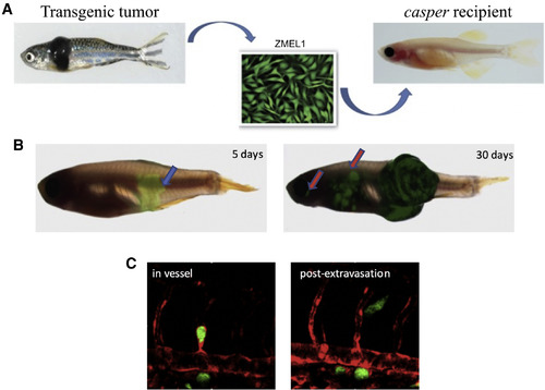
The ZMEL/Casper Transplant Assay Allows for Single-Cell Resolution of the Entire Metastatic Cascade (A) The ZMEL1 line is derived from a transgenic zebrafish melanoma. It is then transplanted into the skin of the transparent casper recipient. (B) Tumors initiate at a defined spot where cells are injected (blue arrow) and metastases (red arrows) can then be followed over time. (C) An example of a single extravasating ZMEL1-GFP cell at a distant site show its appearance while still in the blood vessel (marked by an flk-RFP transgene) and immediately after extravasation out of the blood vessel. |
Reprinted from Developmental Cell, 49, Cagan, R.L., Zon, L.I., White, R.M., Modeling Cancer with Flies and Fish, 317-324, Copyright (2019) with permission from Elsevier. Full text @ Dev. Cell