
- Title
-
The HMG box transcription factors Sox1a and b specify a new class of glycinergic interneurons in the spinal cord of zebrafish embryos
- Authors
- Gerber, V., Yang, L., Takamiya, M., Ribes, V., Gourain, V., Peravali, R., Stegmaier, J., Mikut, R., Reischl, M., Ferg, M., Rastegar, S., Strähle, U.
- Source
- Full text @ Development

sox1a and sox1b are co-expressed in the zebrafish spinal cord. (A,B) Embryos at 24 hpf hybridized to sox1a (A) and sox1b (B) probes. Both genes are expressed in the forebrain, hindbrain and lens (arrowheads), in the otic vesicle (arrows), and in cells along the spinal cord. In addition, sox1a is strongly expressed in the lateral line primordium (asterisk). (C-C″) Fluorescence in situ hybridization (FISH) for sox1a and sox1b mRNA at 24 hpf shows co-expression in V2 (white arrows) and KA neurons. (D) Cells were counted over the yolk extension in a five-somite-long segment. Data are mean±s.d. In the V2 domain, 83±2% of cells (n=103) co-express sox1a and sox1b mRNA, 15±3% of cells express only sox1a, and 2±1% of cells express only sox1b. Similar counts were obtained for KA′ (sox1a/b, 99±4%; sox1a, 0%; sox1b, 1±0%, n=86) or KA″(sox1a/b, 87±3%; sox1a, 1±0%; sox1b, 12±3%, n=115). Six embryos from two independent experiments were counted. (E) Ventral spinal cord domains V1 to V3 with the locations of sox1a+ and sox1b+ KA′ and KA″ neurons (orange circles), and neurons in the V2 domain (V2s, arrow) indicated. Embryos are at 24 hpf. Dorsal is upwards; anterior is leftwards. Scale bars: 200 μm in A,B; 25 μm in C-C″. EXPRESSION / LABELING:
|
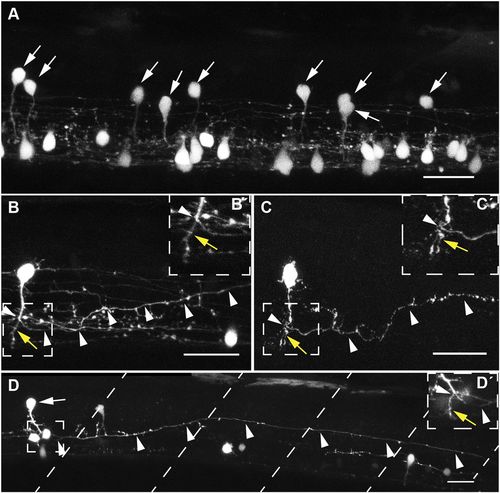
Morphology of V2s neurons. (A) Spinal cord of a sox1a:eGFP embryo with eGFP+ neurons in the V2 domain (arrows) at 60 hpf over a section of three somites above the yolk extension. Ventrally located sox1a:eGFP+ cells without arrows are KA′ and KA″ neurons. sox1a:eGFP+ V2 neurons show an oval-shaped soma at an intermediate spinal cord position and an axon extending ventrally towards the floor plate. (B,B′) sox1a:eGFP+ V2 neuron in a mosaic eGFP knockout embryo at 60 hpf. The ventrally extending axon branches over the floorplate into a long axon descending ipsilaterally (arrowheads) and a short axon branch ending ventrally (B′, yellow arrow). (C,C′) A sox1a+ neuron labelled transiently by TgBAC(sox1a:eGFP) at 48 hpf with an oval-shaped soma and ventrally extending axon that branches into a short axon ending ventrally (C′, yellow arrow) and a long axon descending and rising to an intermediate DV level of the spinal cord (arrowheads). (D,D′) The sox1a:eGFP+ V2 neuron (arrow) extends a long axon ipsilaterally over five somites (somite boundaries are indicated by dashed lines). (D′) Different focal plane showing the short axon branch (yellow arrow) ending ventrally and the main axon branching and descending (arrowheads). Dorsal is upwards; anterior is leftwards. Data are derived from at least two independent experiments. Scale bars: 25 µm in A-C; 100 µm in D. EXPRESSION / LABELING:
|
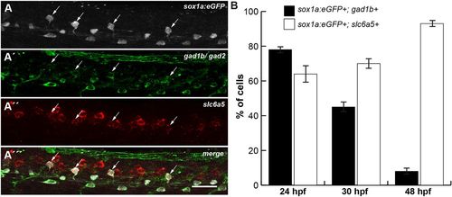
V2s cells develop into glycinergic interneurons. (A-A‴) Multicolour-labelling of a 24 hpf sox1a:eGFP transgenic embryo using FISH with a slc6a5 probe (red), and immunohistochemistry (IHC) with anti-eGFP (white) and anti-Gad1b/Gad2 (green) antibodies. Many sox1a:eGFP+ cells in the V2 domain are both GABA- and glycinergic at 24 hpf (arrows). (B) Percentage of cells expressing sox1a:eGFP and gad1b (black) or sox1a:eGFP and slc6a5 (white) calculated from FISH with a gad1b or slc6a5 probe, and IHC with anti-eGFP antibody at 24, 30 and 48 hpf (Fig. S4). At 24 hpf, 78% of sox1a:eGFP+ cells are GABAergic (n=91 of 117) and 64% are glycinergic (n=34 of 53). At 30 hpf, 45% of sox1a:eGFP+ cells (n=51 of 114) are GABAergic and 70% are glycinergic (n=56 of 80). At 48 hpf, only a minor fraction (8%) still expresses gad1b (n=8 of 98). The majority of sox1a:eGFP cells (93%) have turned on the glycinergic marker by 48 hpf (n=84 of 90). Counts of five to eight embryos from two independent experiments. Data are mean±s.d. Views of spinal cord over yolk extension: dorsal is upwards; anterior is leftwards. Scale bar: 25 µm. |

V2s neurons depend on Notch signalling. (A) Mean number of cells expressing sox1a and gata2a mRNA at 24 hpf (black) and 30 hpf (white) determined by FISH (Fig. S5A-B″). Cells expressing sox1a and gata2a mRNA decreased by about 48% from 24 (black, n=71) to 30 hpf (white, n=34), whereas sox1a:eGFP+ V2 cells co-expressed nkx1.2lb mRNA (Fig. S5C-D″) at similar levels at both stages. (B-E‴) Examples of co-expression of vsx1:GFP, gata2a and sox1amRNA. (B-B‴) A pair of vsx1:GFP+ cells with one cell being vsx1:GFP+;gata2a+;sox1a+ (arrows) extending axons at 24 hpf. Committed V2a cells express only vsx1:GFP (arrowheads). (C-C‴) Example of a vsx1:GFP+ cell co-expressing gata2a (+) at 24 hpf. (D-D‴) A V2s cell that is still vsx1:GFP+ and expresses sox1a mRNA (x) at 26 hpf. (E-E‴) sox1a is co-expressed with gata2a in one cell of a V2a/V2b,s pair (arrows) at 22 hpf. Arrowheads indicate vsx1:GFP+ V2a neurons. (F) Disruption of Notch signalling in sox1a:eGFP embryos from 16 to 24 hpf and assessment of neurotransmitter type at 30 hpf shows a 2.3-fold increase in sox1a:eGFP+ neurons expressing the GABAergic marker gad1b. In contrast, blocking of Notch signalling leads to a 2.3-fold decrease in sox1a:eGFP+;slc6a5+ neurons (DMSO-treated control, white; LY treated, black; for original data, see Fig. S5E-H″). (G-J) sox1a precursor cells divide largely before 24 hpf. sox1a:eGFP+ (green) embryos were treated with EdU (magenta) during two different time windows before processing with EdU click-chemistry at 30 hpf (G). sox1a:eGFP embryos treated from 16 to 24 hpf (H-H″) or from 24 to 30 hpf (I-I″) showing a five-somite spinal cord segment at 30 hpf (sox1a:eGFP+; EdU+ cells, arrows; sox1a:eGFP+; EdU− cells, +). (J) Percentages of EdU+and EdU−; sox1a:eGFP+ cells. Most cells divided before 24 hpf. (A,F) Counts of six to nine embryos from two independent experiments in the V2 domain of the spinal cord above the yolk extension over a five-somite distance. Dorsal is upwards; anterior is leftwards. Data are mean±s.e.m. (A,F) or mean±s.d. (J). Statistical significance was assessed using the unpaired two-tailed Student's t-test. **P≤0.01. Scale bars: 25 µm. EXPRESSION / LABELING:
|

Gata2a and Gata3 are required for expression of sox1a and sox1b in V2 interneurons. (A-E′) Embryos injected with mismatch morpholinos (control, A-E) or with a mixture of morpholinos directed against gata2a and gata3 mRNA (A′-E′). Cells in the V2 domain are indicated by asterisks, KA′ cells by arrowheads and KA″ cells by arrows. (A-A″) Loss of function of Gata2a and Gata3 resulted in a reduction of tal2-expressing KA″ cells, an elimination of KA′ and decrease of tal2+ V2b cells. (B-B″) Reduction of gad67-expressing V2b and KA″ cells was noted in morpholino-injected embryos, whereas loss of function of Gata2a and Gata3 almost abolished gad67+ KA′ cells. (C-C″) Knockdown of Gata2a and Gata3 decreased sox1a+ V2 cells by 25% (n=650 cells) and almost abolished sox1a-expressing KA′. (D-D″) Loss of function of Gata2a and Gata3 decreased sox1b+ V2 cells by 35% (n=2016 cells) and almost eliminated sox1b-expressing KA′. (E-E″) V2a cells (vsx2+) were not affected in knockdown embryos. Data are mean±s.e.m. from 22 to 42 embryos from at least two independent experiments. Cells were counted from the yolk extension to the tail on both sides of the spinal cord. Statistical significance was assessed using the unpaired two-tailed Student's t-test. ***P≤0.001. Scale bar: 25 µm. |

Knockout of sox1a and sox1b causes increase of V2b cells. (A-A″) Knockout of sox1aand sox1b increases gata2a+ cells in the V2 domain by 22%. (B-B″) tal1+ cells are increased by 83% in sox1a−/−; sox1b−/− embryos. (C-C″) Lack of sox1a/b results in an increase of tal2+ cells by 75%. (D-D″) The sox1a/b mutant has 35% more gata3+ cells. (E-E″) vsx2+ cells are not affected by the mutations. (F-F″) At 30 hpf, the gata3+ cells in the V2 domain are increased by 2.3-fold. (G-I) Double FISH of nkx1.2lb (red) and slc6a5 (green) at 30 hpf and quantification of nkx1.2lb+; slc6a5+ cells in I. Glycinergic nkx1.2lb+ cells were decreased by 22% in the mutant. Asterisks indicate double-labelled cells. Counts of gata2a+ cells (A-A″) were derived from the fourth somite to the tail on both spinal cord sides from five embryos. Counts of gata3-, tal2-, tal1-, vsx2- and nkx1.2lb; slc6a5-expressing cells were derived from the spinal cord above the yolk extension over five somites from three to eight embryos (B-F″). V2 cells, white arrowheads (A-F′). Wild-type (white) and mutant (black) values are presented as mean±s.e.m. in A″-F″,I from at least two independent experiments. Statistical significance was assessed using the unpaired two-tailed Student's t-test. *P≤0.05, **P≤0.01, ***P≤0.001. Lateral views at 24 hpf (A-E′) and 30 hpf (F-H″). Dorsal is upwards; anterior is leftwards. Scale bars: 25 µm. |

ZFIN is incorporating published figure images and captions as part of an ongoing project. Figures from some publications have not yet been curated, or are not available for display because of copyright restrictions. PHENOTYPE:
|
|
ZFIN is incorporating published figure images and captions as part of an ongoing project. Figures from some publications have not yet been curated, or are not available for display because of copyright restrictions. EXPRESSION / LABELING:
|