- Title
-
Rab33a and Rab33ba mediate the outgrowth of forebrain commissural axons in the zebrafish brain
- Authors
- Huang, L., Urasaki, A., Inagaki, N.
- Source
- Full text @ Sci. Rep.
|
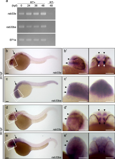
Expression of zebrafish rab33a and rab33ba. (a) RT-PCR analysis of rab33a and rab33ba transcripts. Elongation factor 1a (EF1a) was used as a control. Developmental stages are denoted as hours post-fertilization (hpf). RT-PCR products produced in the presence (RT+) or absence (RT−) of reverse transcriptase were electrophoresed on 2% agarose gels. (b,c) Whole-mount in situ hybridization of rab33a (b) and rab33ba (c) at 24 hpf; (b′) and (c′) show the enlarged lateral (left) and ventral (right) views of the areas indicated by the rectangles. (d,e) Whole-mount in situ hybridization of rab33a (d) and rab33ba(e) at 36 hpf; (d′) and (e′) show the enlarged lateral (left) and ventral (right) views of the areas indicated by the rectangles. Arrowheads and arrows in (b′–e′) indicate the DRC and VRC, respectively. Arrows in (b–e) indicate the hindbrain. See the negative control data for whole-mount in situ hybridization (Supplementary Fig. S3). Scale bars: 100 μm.
|
|
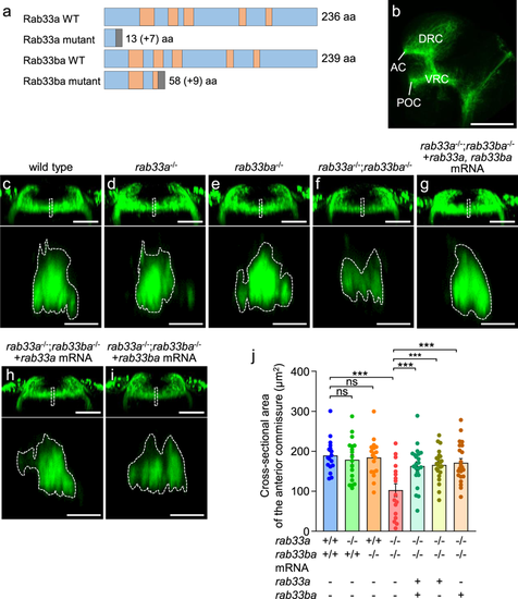
rab33a;rab33ba double mutants display a reduced cross-sectional area of the anterior commissure. (a) Schematic structures of Rab33a and Rab33ba proteins in wild-type and single mutant fish. Frameshift mutations in rab33a and rab33ba result in premature stop codons after aa positions 13 and 58, respectively. The gray boxes indicate amino acids added by the frameshift mutations, and the numbers in brackets indicate the numbers of these additional residues. The regions involved in GTP/GDP-binding and GTPase activity11,17 are indicated by the orange color. (b) A representative lateral view of a 36 hpf wild-type zebrafish brain immunolabeled with anti-acetylated tubulin antibody. See Movie 1. Abbreviations: AC, anterior commissure; POC, postoptic commissure. Scale bar: 100 μm. (c–i) Frontal views (upper panels) of the anterior commissures of wild-type control (c), rab33a−/− single mutant (d), rab33ba−/− single mutant (e) and rab33a−/−;rab33ba−/−double mutant (f) embryos at 36 hpf. In (g), rab33aand rab33ba mRNAs were injected into a rab33a−/−;rab33ba−/− double mutant embryo for rescue analysis. In (h,i) rab33a mRNA (h) and rab33ba mRNA (i) were injected into a rab33a−/−;rab33ba−/− double mutant embryo for rescue analysis. Scale bars: 50 µm. The lower panels show cross-sections at the dotted lines shown in the frontal views. Scale bars: 10 µm. (j) The cross-sectional area of the anterior commissure obtained from the data analyses in (c–i, lower panels). Wild-type control (n = 17), rab33a−/−single mutant (n = 18), rab33ba−/− single mutant (n = 18) and rab33a−/−;rab33ba−/− double mutant (n = 17) embryos, and rab33a−/−;rab33ba−/− double mutant embryos with rab33a and rab33ba mRNAs (n = 22), rab33a mRNA (n = 22) and rab33ba mRNA (n = 22) were analyzed at 36 hpf. Data are mean ± SEM; ***P < 0.01; ns, not significant (one-way ANOVA with Tukey’s post hoc test).
PHENOTYPE:
|
|
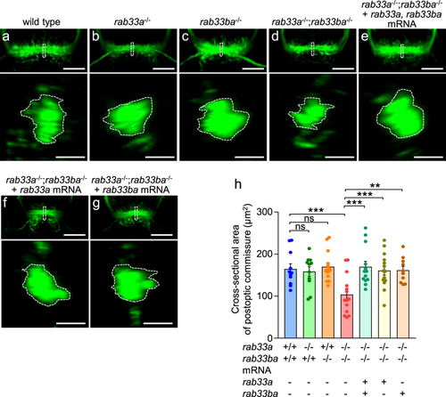
rab33a;rab33ba double mutants display a reduced cross-sectional area of the postoptic commissure. (a–g) Representative frontal views (upper panels) of the postoptic commissures of wild-type control (a), rab33a−/− single mutant (b), rab33ba−/− single mutant (c) and rab33a−/−;rab33ba−/− double mutant (d) embryos at 36 hpf. In (e), rab33a and rab33ba mRNAs were injected into a rab33a−/−;rab33ba−/− double mutant embryo for rescue analysis. In (f,g), rab33a mRNA (f) or rab33ba mRNA (g) were injected into a rab33a−/−;rab33ba−/− double mutant embryo for rescue analysis. Scale bars: 50 µm. The lower panels show the cross-sections at the dotted lines shown in the frontal views. Scale bars: 10 µm. (h) The cross-sectional area of the postoptic commissure obtained from the data analyses in (a–g; lower panels). Wild-type control (n = 11), rab33a−/− single mutant (n = 11), rab33ba−/− single mutant (n = 14) and rab33a−/−;rab33ba−/− double mutant (n = 14) embryos, and rab33a−/−;rab33ba−/− double mutant embryos with rab33a and rab33ba mRNAs (n = 13), rab33a mRNA (n = 14) and rab33ba mRNA (n = 10) were analyzed at 36 hpf. Data are mean ± SEM; ***P < 0.01; **P < 0.02; ns, not significant (one-way ANOVA with Tukey’s post hoc test).
PHENOTYPE:
|
|
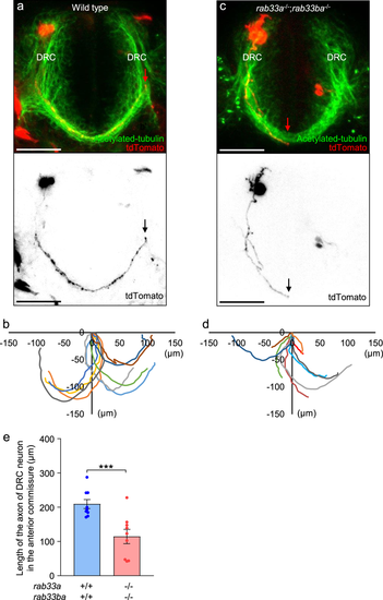
The rab33a;rab33ba double mutation inhibits axonal extension in the anterior commissure. (a,c) Representative dorsal views (upper panels) of the anterior commissures immunolabeled with anti-acetylated tubulin antibody and individual DRC neurons expressing tdTomato in wild-type control (a) and rab33a−/−;rab33ba−/− double mutant (c) embryos at 36 hpf. The lower panels show only the tdTomato-labeled neurons in the upper panels. The tips of the axons are indicated by arrows. Scale bars: 50 µm. (b,d) The trajectories of individual axons of the tdTomato-labeled DRC neurons in wild-type control (b) and rab33a−/−;rab33ba−/− double mutant (e) embryos at 36 hpf. The cell body positions are normalized at (x = 0 μm, y = 0 μm). (e) The length of the axon of DRC neurons in the anterior commissure obtained from the data analyses in (b,d). Axons of wild-type control (n = 9) and rab33a−/−;rab33ba−/−double mutant (n = 9) neurons were analyzed at 36 hpf. Data are expressed as mean ± SEM; ***P < 0.01 (unpaired Student’s t test).
|


