
- Title
-
Female Sex Development and Reproductive Duct Formation Depend on Wnt4a in Zebrafish
- Authors
- Kossack, M.E., High, S.K., Hopton, R.E., Yan, Y.L., Postlethwait, J.H., Draper, B.W.
- Source
- Full text @ Genetics

ZFIN is incorporating published figure images and captions as part of an ongoing project. Figures from some publications have not yet been curated, or are not available for display because of copyright restrictions. EXPRESSION / LABELING:
|
|
ZFIN is incorporating published figure images and captions as part of an ongoing project. Figures from some publications have not yet been curated, or are not available for display because of copyright restrictions. EXPRESSION / LABELING:
PHENOTYPE:
|
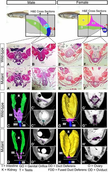
Duct morphology in wnt4a mutant and wild-type males. From the posterior end of each testis, wild-type males developed duct deferens (A) that joined to form the fused duct deferens (B). Mutant males, however, failed to form a full duct deferens (A′) or a fused duct deferens (B′). Three-dimensional renderings built from individual traces of sections (C′, C″, D′, and D″) of (C) the wild-type ducts and (D) mutant ducts show that the mutant ducts never fully connected to the genital orifice. H&E-stained sections of wild-type females showed an oviduct that wrapped around the posterior of the ovaries (E, G, and G′) and extended ventroposteriorly (F, G, and G″) out to the genital orifice. Mutant females, however, failed to organize an oviduct around the ovary (E′, H, and H′) and did not form a connective duct to the genital orifice (F′, H, and H″). See movies in Files S1 and S2. Bar, 100 µm. I, intestine; K, kidney; T, testis, O, ovary; OD, oviduct; GO, genital orifice; DD, duct deferens; FDD fused duct deferens. |
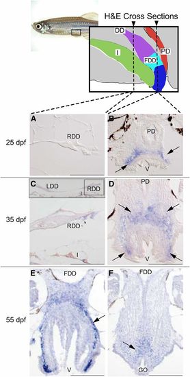
Expression of wnt4a during reproductive duct development in zebrafish wild-type AB strain males. Alternate serial cross sections of wild-type males were stained by H&E to follow duct growth (see Figure S6) or prepared for in situ hybridization to reveal wnt4a expression, shown here. (A) Expression analysis showed that at 25 dpf, the duct deferens (cross section of the right duct shown here) lacked wnt4a expression. (B) At 25 dpf, however, wnt4a expression (→’s) appeared dorsal to the posterior vent, just anterior to the eventual connection of the fused duct deferens and the genital orifice. (C) At 35 dpf, the duct deferens continued to show little wnt4a expression. Insert shows the left and right duct deferens dorsal to the intestine. (D) At 35 dpf, wnt4a expression (→’s) appeared dorsal and lateral to the posterior vent, just anterior to the eventual connection of the fused ducti deferens and the genital orifice. (E) At 55 dpf, in a section just anterior to the fusion of the duct deferens to the vent, wnt4a expression (→) appeared dorsal to the posterior vent, just anterior to the connection of the fused ducti deferens and genital orifice. (F) At 55 dpf, in a section at the level of the connection of the fused duct and the vent, wnt4a expression (→) appeared just dorsal to the genital orifice. DD, ducti deferens; FDD, fused ducti deferens; GO, genital orifice; I, intestine; LDD, left duct deferens; PD, pronephric duct; RDD, right duct deferens; V, vent. Bar, 100 µm. |
|
Unillustrated author statements PHENOTYPE:
|