
- Title
-
A missense variant in SLC39A8 is associated with severe idiopathic scoliosis
- Authors
- Haller, G., McCall, K., Jenkitkasemwong, S., Sadler, B., Antunes, L., Nikolov, M., Whittle, J., Upshaw, Z., Shin, J., Baschal, E., Cruchaga, C., Harms, M., Raggio, C., Morcuende, J.A., Giampietro, P., Miller, N.H., Wise, C., Gray, R.S., Solnica-Krezel, L., Knutson, M., Dobbs, M.B., Gurnett, C.A.
- Source
- Full text @ Nat. Commun.

Skeletal abnormalities and growth deficit in slc39a8−/− zebrafish. a Thoracic and caudal vertebral malformations causing mild spinal curvature in slc39a8−/− zebrafish (bottom) compared to WT (top) stained with alizarin red. Magnified images show vertebral fusions in KO fish. Scale bar 5 mm. b slc39a8−/− zebrafish have more spinal abnormalities than WT fish. c Reduced length of slc39a8−/− zebrafish (N = 20) compared to wild-type (N = 20) at 9 mpf ***p < 10−6, t-test. Lines are mean, first and third quartiles PHENOTYPE:
|

ZFIN is incorporating published figure images and captions as part of an ongoing project. Figures from some publications have not yet been curated, or are not available for display because of copyright restrictions. PHENOTYPE:
|
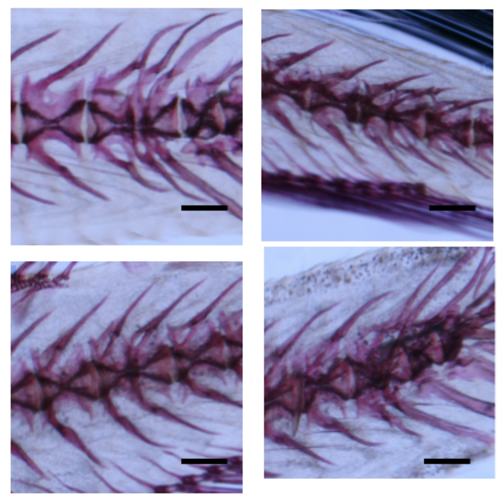
Additional Images of abnormal slc39a8 KO zebrafish after alizarin red staining. Various skeletal abnormalities were observed among slc39a8 KO zebrafish including fused vertebrae and abnormal bone growth along the spine. Scale bar 1mm. PHENOTYPE:
|
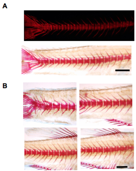
Additional Images of abnormal slc39a8 KO zebrafish after alizarin red staining at 12 weeks post-fertilization. (A) A single mutant slc39a8 zebrafish demonstrating spinal fusion by fluorescent or light microscopy. (B) Various skeletal abnormalities were observed among slc39a8 KO zebrafish including fused vertebrae and abnormal bone growth along the spine (4 different fish shown). Shown are homozygous mutant slc39a8 zebrafish produced from a cross between two heterozygous mutant slc39a8 zebrafish and genotyped for the mutation. Scale bar 2mm. PHENOTYPE:
|

Calcein staining of slc39a8 mutant zebrafish at 13 dpf. Three example -/- slc39a8 zebrafish are shown. Individual zebrafish were stained with calcein and imaged for fluorescence to look for early skeletal developmental abnormalities. Scale bar 0.5 mm. PHENOTYPE:
|
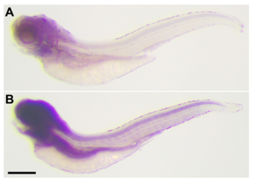
Expression of slc39a8 mRNA revealed by whole-mount in situ hybridization. Lateral view of 4 days post-fertilization (dpf) zebrafish embryos. (A) Control with sense probe indicating background level of staining. (B) Detection of slc39a8 mRNA using anti-sense probe. Slc39a8 is expressed in the eye, brain, spinal cord and endodermal tissues. Scale bar: 0.2 mm. |