
- Title
-
Prednisolone induces osteoporosis-like phenotypes via focal adhesion signaling pathway in zebrafish larvae
- Authors
- Huo, L., Wang, L., Yang, Z., Li, P., Geng, D., Xu, Y.
- Source
- Full text @ Biol. Open
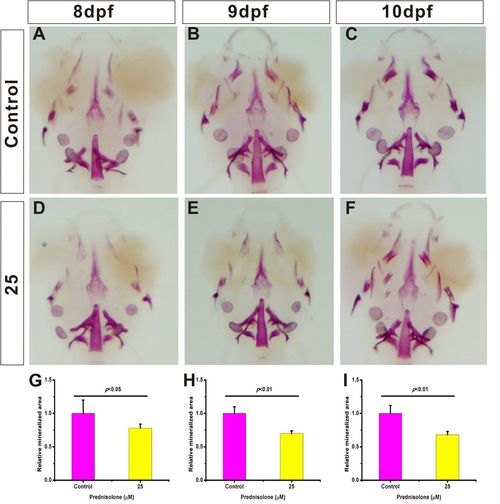
Establishment of a zebrafish larva GIOP model using 25 μM prednisolone. (A-F) 8−10 dpf zebrafish treated with 25 μM prednisolone. Whole mount skeletal staining was performed on fixed tissues. The mineralized tissue is stained purple and all other tissues are transparent. (G-I) Digital image analysis of stained area and staining density by assessment of fluorescence intensity. The stained mineralized tissue was quantified using analysis software. Mean values are plotted (n=10) and the Student's t-test was performed to determine statistical significance. PHENOTYPE:
|
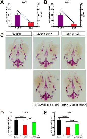
Osteoporosis-like phenotypes caused by prednisolone through itga10 and itgbl1. (A) qRT-PCR confirmed that itga10 expression is down-regulated by prednisolone. (B) qRT-PCR confirmed that itgbl1 expression is down-regulated by prednisolone. (C) The gRNAs for itga10 and itgbl1 were designed and injected into single-cell embryos. Whole mount skeletal staining was performed on fixed tissues. (D,E) Digital image analysis of stained area, and staining density by fluorescence intensity measurement. The stained mineralized tissue was quantified using analysis software. Mean values are plotted (n=5) and the Student’s t-test was performed to determine statistical significance. |
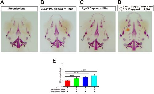
Exogenous itga10 and itgbl1 capped mRNA rescue the osteoporosis-like phenotype of prednisolone. (A) After treatment with 25 µM prednisolone, whole mount skeletal staining was performed at 10 dpf. (B) itga10 capped mRNA was injected into single-cell embryos, larvae were treated with 25 μΜ prednisolone at 6 dpf and whole mount skeletal staining was conducted at 10 dpf. (C) itgbl1 capped mRNA was injected into single-cell embryos. Larvae were treated with 25 μΜ prednisolone at 6 dpf, and whole mount skeletal staining was conducted at 10 dpf. (D) itga10 and itgbl1 capped mRNAs were co-injected into single-cell embryos, larvae were treated with 25 μΜ prednisolone at 6 dpf, and whole mount skeletal staining was conducted at 10 dpf. (E) Digital image analysis of stained area and staining density by fluorescence intensity measurement, and the stained mineralized tissue was quantified using analysis software. Mean values are plotted (n=5) and the Student's t-test was performed to determine statistical significance. |
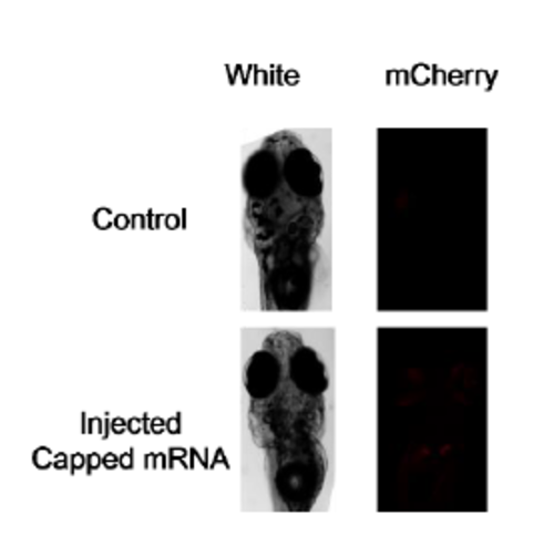
Capped mRNA was partly worked in the 10dpf larva fish. The itga10 and itgbl1 fused mCherry was injected eggs. We can founded weak but obvious mCherry fluorescence in the injected 10dpf fish. |