- Title
-
Human MLL-AF9 Overexpression Induces Aberrant Hematopoietic Expansion in Zebrafish
- Authors
- Tan, J., Zhao, L., Wang, G., Li, T., Li, D., Xu, Q., Chen, X., Shang, Z., Wang, J., Zhou, J.
- Source
- Full text @ Biomed Res. Int.
|
MLL-AF9 overexpression in zebrafish embryos and activation of MLL downstream genes. MLL-AF9 mRNA was injected into zebrafish at the 1–2-cell stage (around 5ng per embryo). Injected embryos and control embryos were harvested at 48hpf. (a) Western blotting of the N-terminus of the MLL protein in MLL-AF9-injected and control embryos. (b) Relative expression of MLL downstream genes analyzed by qRT-PCR. (c–e) WISH assays of hoxa9a, hoxb5a, and meis1 in MLL-AF9-injected and control embryos at 24hpf. (f, g) Morphology of the hematopoietic cells of control embryos and MLL-AF9-injected embryos at 48 hpf. |
|
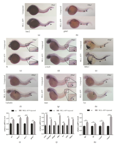
Induction of zebrafish primitive and definitive hematopoiesis by MLL-AF9. WISH assays and qRT-PCR analysis of hematopoietic markers in MLL-AF9-injected and control embryos. (a–c) WISH assays of hematopoietic markers (lmo2 and gata1) and a vasculature formation marker (fli) in MLL-AF9-injected and control embryos at 24hpf. (d–h) WISH assays at 48hpf of the definitive hematopoietic stem cell marker c-myb, the erythropoietic marker hbbe1, and the myeloid lineage markers l-plastin, mpy, and lyz. Relative expression of the marker genes for (i) primitive hematopoiesis at 24hpf, (j) hematopoiesis at 48hpf, (k) vasculature formation (flk, fli) at 24hpf, and (k) definitive hematopoiesis initiation (c-myb and runx1) at 48hpf analyzed by qRT-PCR. |
|
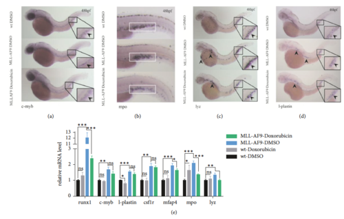
Doxorubicin inhibition of MLL-AF9-induced hematopoiesis. Doxorubicin and DMSO (control) were administered to MLL-AF9-injected and wild-type embryos from 24 to 48hpf. WISH assays of the (a) definitive hematopoietic stem cell marker c-myb and (b–d) myeloid lineage markers mpo, lyz, and l-plastin in wild-type embryos treated with DMSO (top), MLL-AF9-injected embryos treated with DMSO (middle), and MLL-AF9-injected embryos treated with doxorubicin (bottom) at 48hpf. (e) Relative expression of the marker genes for definitive hematopoiesis (runx1 and c-myb) and myeloid hematopoiesis (l-plastin, csf1r, mfap4, mpo, and lyz) in wild-type and MLL-AF9-injected embryos treated with DMSO or doxorubicin analyzed by qRT-PCR. Wild-type embryos treated with DMSO were used as a control. |
|
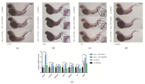
Amelioration of MLL-AF9-induced hematopoiesis by MI-2. The menin inhibitor MI-2 (2.5mmol/L) and DMSO (control) were administered to MLL-AF9-injected and wild-type embryos from 24 to 72hpf. WISH assays of the (a) definitive hematopoietic stem cell marker c-myb and (b–d) myeloid lineage markers mpo, lyz, and l-plastin in wild-type embryos treated with DMSO (top), MLL-AF9-injected embryos treated with DMSO (middle), and MLL-AF9-injected embryos treated with MI-2 (bottom) at 72hpf. (e) Relative expression of the marker genes for definitive hematopoiesis (runx1 and c-myb) and myeloid hematopoiesis (l-plastin, csf1r, mfap4, mpo, and lyz) in wild-type and MLL-AF9-injected embryos treated with MI-2 or doxorubicin analyzed by qRT-PCR. Wild-type embryos treated with DMSO were used as a control. |
|
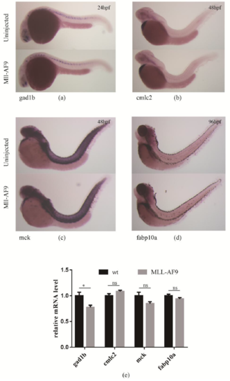
Effect of MLL-AF9 on non-hematopoietic tissue markers. WISH assays of non-hematopoietic markers, including (a) the neural marker gad1b at 24hpf, (b) the cardiac chamber marker cmlc2 at 48hpf, (c) the skeletal muscle marker mck at 48hpf, and (d) the hepatocyte marker fabp10a at 96hpf in MLL-AF9-injected and control embryos. (e) qRT-PCR analysis of gad1b, cmlc2, mck, and fabp10a at 24, 48, 48, and 96hpf, respectively. |


