- Title
-
A High-Content Screen Reveals New Small-Molecule Enhancers of Ras/Mapk Signaling as Probes for Zebrafish Heart Development
- Authors
- Saydmohammed, M., Vollmer, L.L., Onuoha, E.O., Maskrey, T.S., Gibson, G., Watkins, S.C., Wipf, P., Vogt, A., Tsang, M.
- Source
- Full text @ Molecules
|
In vivo whole organism chemical screen identified new hyperactivators of Fgf/Ras/Mapk signaling. (A) Representative fluorescence micrographs of Tg(dusp6:EGFP) embryos (30 hpf) treated at 24 hpf for 6 h. Top panel, original scan image; bottom panel, images with Cognition Network Technology (CNT) algorithm applied. All of the compounds listed above images were at 10 µM. (B) Confirmatory dose response assay using repurchased small molecules: ST06994, ST011282, and ST020101. Each data point represents an independent assessment of total green fluorescent protein (GFP) intensity in the head of zebrafish larvae (n = 8). Horizontal bars denote the mean ± S.D. from two or three independent experiments. |
|
ST006994, ST020101, and ST011282 treatment induced the dorsalization and expansion of cardiac progenitor cells in zebrafish, which is a Fgf hyperactivation phenotype. (A) Semi-quantitative RT-PCR showing an increase of dusp6 and EGFP transcripts after chemical treatment. (B) Representative images of whole mount in situ hybridization showing tbxta, chordin, and pax2a/egr2b expression after compound treatment. tbxta expression was increased in the tailbud (blue arrow), and chordin expression was expanded in the dorsal region when compared with DMSO control (distance between two red arrows). The neural markers pax2a and egr2b showed lateral expansion (red brackets), which is indicative of dorsalized phenotype from increased early Fgf activity. |
|
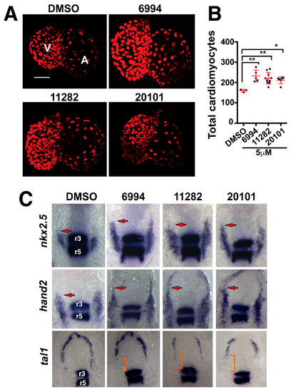
Fgf hyperactivators regulate heart size by increasing cardiac progenitor populations. (A) Fluorescence micrographs of cardiomyocyte nuclei from 72 hpf transgenic larvae treated for somite stages one through eight with 5 µM BCI, ST006994, ST011282, or ST020101. Images are representative for each compound. (B) The quantification of cardiomyocytes after small molecule treatment. All three compounds significantly increased the numbers of cardiomyocytes in the developing zebrafish heart. Each point represents a single larvae. (C) Whole mount in situ hybridization shows the expression of cardiac progenitor markers (nkx2.5 and hand2) and endothelial progenitors (tal1) after compound treatment. egr2b was used to mark the hindbrain rhombomeres 3 and 5 (r3 and r5). Arrows mark the rostral most domain of the cardiac progenitor population. Brackets mark the region of decreased endothelial tal1 expression. |


