- Title
-
Investigation of Islet2a function in zebrafish embryos: Mutants and morphants differ in morphologic phenotypes and gene expression
- Authors
- Moreno, R.L., Williams, K., Jones, K.L., Ribera, A.B.
- Source
- Full text @ PLoS One
|
The islet2a TALEN mutant. (A) The sequence of homozygous islet2a105 genomic DNA revealed a 13-nuc deletion (at position *) that removed a restriction enzyme site and introduced a frameshift. (B) Schematic of wildtype and mutant Islet2a proteins. The islet2a105 sequence predicts a truncated Islet2a protein product lacking the homeobox (HD) and LIM2 (L2) domains and containing only a portion (~70%) of the LIM1 (L1) domain. The Isl11/2 monoclonal antibody used in this study was raised against a carboxy-terminus sequence not present in the predicted mutant protein. (C-F) Expression of islet2a mRNA (C, D) and Isl1/2 immunoreactivity (E, F) in wildtype (wt) and homozygous mutant (mut) islet2a 28 hpf embryos. Images are lateral views of embryos, with dorsal up and anterior to the left. Bright field and fluorescent images have been merged. (C, D) A tissue that expresses islet2a, but not islet1 or islet 2b, was used to assess the efficacy of the TALEN mutation. The proctodeum (P), just caudal to the posterior end of the yolk sac extension (yse) emerges during embryonic stages and develops later into the anal passage. In both wildtype (n = 6) and mutant (n = 8) embryos, the proctodeum expressed islet2a mRNA. (E, F) The Isl1/2 antibody immunolabeled the proctodeum in wildtype (E; n = 10) but not mutant (F; n = 7) 28 hpf embryos. Scale bar in F, for C-F: 25 μm. |
|
Islet2a morphant but not mutant embryos displayed altered CaP morphology. (A-E) In live tg(mnx1:gfp) 24 hpf embryos, CaP neurons expressed gfp in their somas and axons. (A, B) Injection of a T-MO (MO) led to truncation of ventrally projecting axons (asterisks, B) compared to control (Ctl, A), as previously reported [15, 17]. (C-E) In wildtype (wt, C), heterozygous (het, D) and mutant (mut, E) islet2a embryos, CaP neuron axon growth and trajectories appeared normal regardless of genotype. Sample size ranged from 8–30 per condition. Scale bar in E, for A-E: 50 μm. (F, G) In fixed non-transgenic 28 hpf embryos, zn1/znp1 immunoreactivity did not reveal any differences in CaP axon morphology between wildtype (F; n = 9) versus mutant (G; n = 9) embryos. |
|
Islet2a morphant, but not mutant, larvae have a reduced number of dSMNs. (A-C) In 72 hpf tg(islet1:gfp) larvae, dSMNs expressed gfp in their somas and axons. In addition, the zn8 antibody recognized the neurolin protein (red), expressed on SMN somas and axons [38]. (A) In uninjected 72 hpf larvae, the majority of zn8+ neurons also expressed gfp. (B) Following injection of the control MO (Ctl MO), zn8+ (red) neurons continued to express gfp. In addition, dSMNs developed normally with respect to axon morphology (arrowhead), as assessed by zn8 immunolabeling (red). (C) Injection of the T-MO led to a decrease in the number of zn8+ neurons that coexpressed gfp. Scale bar in A, for A-C: 50 μm. (D) In tg(islet1:gfp) 72 hpf larvae, the numbers of gfp+ somas were reduced by injection of the Ctl MO (n = 17) and further reduced by injection of the T-MO (n = 20) compared to uninjected embryos (n = 10). ***, p<0.0001; **, p<0.0003; 8, p<0.0006; ANOVA with post-hoc Bonferroni. (E-G) In live 72 hpf tg(islet1:gfp) larvae, dSMNs appeared normal in number and morphology in homozygous mutant (mut; G) compared to heterozygous (het; F) and homozygous wildtype (wt; E) 72 hpf embryos. (H) Cell counts indicated that the number of dSMN somas was not reduced in mutant (n = 9) compared to wildtype (n = 5) or heterozygous (n = 8) islet2a 72 hpf embryos. EXPRESSION / LABELING:
PHENOTYPE:
|

ZFIN is incorporating published figure images and captions as part of an ongoing project. Figures from some publications have not yet been curated, or are not available for display because of copyright restrictions. |
|
CaPs expressed islet2a, but not islet1 or islet2b, in wildtypes and mutants. The expression patterns of islet1 (A [n = 6], B [n = 8], islet2a (C [n = 6], D [n = 8]) and islet2b (E [n = 9], F [n = 8]) in CaPs were examined in 28 hpf sibling wildtype (A, C, E) and islet2a mutant (B, D, F) tg(mnx1:gfp) embryos. At 28 hpf, VaP (variably-present CaP duplicate) is present in some segments. In A-F, white and green asterisks denote lone CaPs and CaP/VaP pairs, respectively. Novel, ectopic expression of either islet1 or islet2b in CaPs was not detected. EXPRESSION / LABELING:
|
|
CaPs expressed reduced levels of Isl1/2 immunoreactivity in islet2a mutant embryos. (A) In 28 hpf tg(mnx1:gfp) wildtype (wt, n = 15) embryos, CaPs (*) expressed both gfp (green) and Isl1/2 immunoreactivity (red), as revealed by the merged yellow signal. (B) The Isl11/2 immunosignal of Panel A is shown separately. In A-D, dotted lines circle the cell bodies of CaP/VaPs that were immunopositive for Isl1/2. In comparison to other ventral neurons, CaP Isl1/2 immunolabeling was more intense. (C) In 28 hpf tg(mnx1:gfp) mutant (mut, n = 14) embryos, CaPs (*) expressed gfp. However, compared to wildtype (A), the CaP Isl1/2 fluorescent immunolabel signal was less intense. Further, other ventral neurons continued to display Isl1/2 immunoreactivity. (D) The Isl1/2 signal of Panel C is viewed separately. Compared to wildtype (B), CaPs expressed reduced levels of Isl1/2 immunoreactivity. Further, despite the weak signal in CaPs, Isl1/2 immunoreactivity was present in other ventral neurons at levels similar to wildtype (B). Scale bar in D for A-D: 25 μm. (E, F) Examination of CaP Isl1/2 immunolabeling dorsal to the proctodeum. (E) In wildtype embryos, both the proctodeum (white arrow) and CaPs (asterisks) displayed Isl1/2 immunolabeling. One CaP, contained within white dotted line box, is shown at higher magnification in the inset. (F) In mutant embryos, Isl1/2 immunolabeling was not detected in the proctodeum, consistent with loss of Islet2a protein expression. Despite this, a low level of Isl1/2 immunoreactivity persisted in CaPs (asterisks; one CaP shown at higher power in inset). |
|
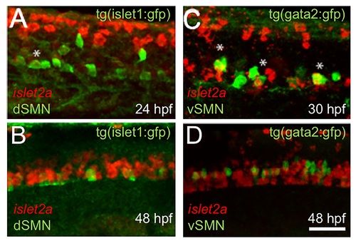
RNA in situ hybridization reveals islet2a expression in a subpopulation of SMNs. (A-D) RNA in situ hybridization was performed using transgenic lines that express gfp in either dSMNs (tg(isl1:gfp); A, B) or vSMNs (tg(gata2:gfp); C, D). The red RNA in situ hybridization signal for islet2a is not detected in gfp+ dorsally-projecting SMNs at either 24 (A) or 48 (B) hpf. In contrast, islet2a RNA is detected in a subset of ventrally projecting SMNs (C, D). Scale bar in D, for A-D: 25 μm. |

