
- Title
-
Assessing the association between hypoxia during craniofacial development and oral clefts
- Authors
- Küchler, E.C., Silva, L.A.D., Nelson-Filho, P., Sabóia, T.M., Rentschler, A.M., Granjeiro, J.M., Oliveira, D., Tannure, P.N., Silva, R.A.D., Antunes, L.S., Tsang, M., Vieira, A.R.
- Source
- Full text @ J Appl Oral Sci
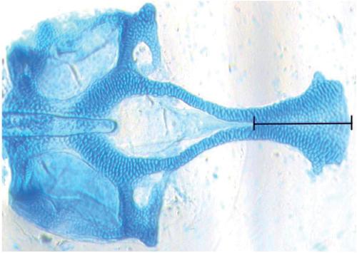
Ventral view. Landmarks used to measure the length of the anterior portion of the ethmoid plate formation |
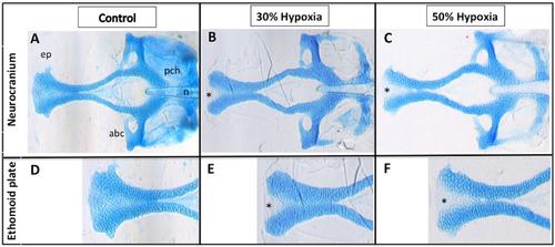
Hypoxia results in craniofacial defects. (A-F) Ventral view of 5 dpf larvae stained with Alcian Blue; (A-C) Dissection of the neurocranium; (D-F) Closer view of the ethmoid plate; (B, C, E and F) Morphological alterations in the anterior area of the ethmoid plate, including a gap in the anterior edge forming a cleft; (C and F) Results of a slightly more severe phenotype in the ethmoid plate (deeper cleft). abc= anterior basicapsular commissure; ep= ethmoid plate; n= notochord; pch= parachordal; * indicates cleft in the ethmoid plate PHENOTYPE:
|