
- Title
-
Knockout of zebrafish interleukin 7 receptor (IL7R) by the CRISPR/Cas9 system delays retinal neurodevelopment
- Authors
- Cai, S., Chen, Y., Shang, Y., Cui, J., Li, Z., Li, Y.
- Source
- Full text @ Cell Death Dis.
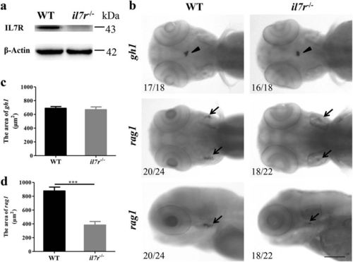
The validation of the il7r−/− mutant by western blotting and whole-mount in situ hybridisation. a The expression of IL7R protein in WT and il7r−/− larvae at 4 dpf. Note that no IL7R protein is detected in il7r−/− larvae. b The images of whole-mount in situ hybridisation with gh1 (arrowheads) and rag1 (arrows) mRNA probes in WT and il7r−/− larvae at 4 dpf. c-d The statistical analysis of gh1-positive area (c) or rag1-positive area (d) between WT and il7r−/− larvae. Note that rag1 expression is significantly decreased in il7r−/− larvae. Results are represented as means ± SEM, ***P < 0.001. The upper four panels in b: dorsal view. The lower two panels in b: dorsal is up, and rostral is left. Scale bar in b: 100 μm EXPRESSION / LABELING:
PHENOTYPE:
|

The phenotypes of the il7r−/− mutant. a-j Phenotypes of WT and il7r−/− embryos at 12 hpf (a and f), 24 hpf (b and g), 48 hpf (c and h) and 72 hpf (d and i). e and j are the magnified images of the heads from d and i, respectively. k Statistical analysis of body length in WT and il7r−/− embryos at 72 hpf. Note that the body length of embryos in the il7r−/− group is shorter than that embryos in the WT group, results are represented as means ± SEM (n = 30), ***P < 0.001. l Statistical analysis of eye size between the two groups. Note that the il7r−/− mutants have smaller eyes, results are represented as means ± SEM (n = 30), **P < 0.01. Dorsal is up, and rostral is left in c, d, e, h, i and j. Scale bar: a-d and f-i, 200 μm; e and j, 100 μm PHENOTYPE:
|

Retinal development following il7r knockout. a-d HE staining and magnified images of retinas from wild-type (WT, a and b) and il7r−/− (c and d) embryos at 72 hpf. e–h Images of Zpr1 or Zpr3 immunofluorescence staining of retinas in WT (e and f) and il7r−/− (g and h) embryos at 72 hpf. i-j Statistical analysis of Zpr1-positive signals (i) and Zpr3-positive signals (j) between WT and il7r−/− retinas. Note that the Zpr1-positive area (i) and Zpr3-positive area (j) are significantly decreased in il7r−/− retinas. Results are represented as means ± SEM (n = 10), **P < 0.01, ***P < 0.001. Scale bar: a and c, 40 μm; b and d, 10 μm; e–h, 50 μm. GCL ganglion cell layer, INL inner nuclear layer, ONL outer nuclear layer, ON optic nerve EXPRESSION / LABELING:
PHENOTYPE:
|

ZFIN is incorporating published figure images and captions as part of an ongoing project. Figures from some publications have not yet been curated, or are not available for display because of copyright restrictions. |
|
ZFIN is incorporating published figure images and captions as part of an ongoing project. Figures from some publications have not yet been curated, or are not available for display because of copyright restrictions. PHENOTYPE:
|

Retinal development following il7r knockout at 60 hpf. (a-d) HE staining and magnified images of retinas from wild-type (WT, a and b) and il7r-/- (c and d) embryos. (e-h) Images of Zpr1 or Zpr3 immunofluorescence staining of retinas in WT (e and f) and il7r-/- (g and h) embryos. (i-j) Statistical analysis of Zpr1-positive signals (i) and Zpr3-positive signals (j) between WT and il7r-/- retinas. Note that the Zpr1-positive area (i) and Zpr3-positive area (j) are significantly decreased in il7r-/- retinas. Results are represented as means ± SEM (n=10), ***P<0.001. Scale bar: (a and c), 40 μm; (b and d), 10 μm; (e-h), 50 μm. Abbreviations: GCL: ganglion cell layer; INL: inner nuclear layer; ONL: outer nuclear layer. |