
- Title
-
Roles of maternal wnt8a transcripts in axis formation in zebrafish
- Authors
- Hino, H., Nakanishi, A., Seki, R., Aoki, T., Yamaha, E., Kawahara, A., Shimizu, T., Hibi, M.
- Source
- Full text @ Dev. Biol.
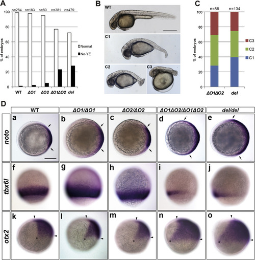
Role of ORF1 and ORF2 in the zygotic wnt8a-mediated development of caudal and ventrolateral tissues. (A) Percentage of mutants showing a lack of yolk extension (no yolk extension: No-YE). Embryos were obtained by crossing WT, or wnt8aΔO1(ΔO1), wnt8aΔO2 (ΔO2), wnt8aΔO1ΔO2 (ΔO1ΔO2), or wnt8adel (del) heterozygote pairs. Phenotypes were observed at 30 hpf. The number of observed embryos is indicated. (B) WT and wnt8a mutant phenotypes. Lateral views with anterior to the left. The No-YE embryos were categorized into three classes. C1 embryos had a slightly shortened and ventrally bent tail, C2 embryos had a short and curled tail, and C3 embryos had an enlarged head and a truncated tail. This phenotype classification is essentially the same as one described in a previous publication (Shimizu et al., 2005), except for being based on the No-YE phenotype. (C) Proportion of No-YE embryos showing each phenotype class. The number of observed embryos is indicated. (D) Expression of noto (formerly called floating head, shield stage, a-e), tbx6l (tbx6, 80% epiboly stage, f-j), and otx2 (100% epiboly stage, k-o) in WT (a, f, k) and ΔO1 (b, g, l), ΔO2 (c, h, m), ΔO1ΔO2 (d, i, n), and del (e, j, o) homozygous mutants. Animal pole views with dorsal to the right (a-e). Lateral views with dorsal to the right (f-o). noto, tbx6l, and otx2 are markers of the axial mesoderm, the paraxial mesoderm, and the rostral neuroectoderm (forebrain and midbrain), respectively. The lateral limits of noto expression are marked by arrows, the rostral and caudal limits of otx2 expression are marked by arrowheads, and the ventral limit of otx2 expression is marked by an asterisk. The noto expression was not greatly affected in the ΔO1 and ΔO2 single mutants, but it was prominently expanded in the ΔO1ΔO2 and del mutants (ΔO1, n=0/86; ΔO2, n=0/94; ΔO1ΔO2, n=10/42; del, n=15/44 in embryos from the heterozygous crosses; ΔO1, n=0/7; ΔO2, n=0/7; ΔO1ΔO2, n=4/4; del, n=6/6 in genotyped homozygotes). The tbx6l expression was not affected in the ΔO1 or ΔO2 mutants, but was reduced or absent in the ΔO1ΔO2 and del mutants (ΔO1, n=0/353; ΔO2, n=0/96; ΔO1ΔO2, n=27/83; del, n=37/140 in embryos from the heterozygous crosses; ΔO1, n=0/22; ΔO2, n=0/9; ΔO1ΔO2, n=11/11; del, n=10/10 in the genotyped homozygotes). The otx2 expression was not affected in the ΔO1 or ΔO2 mutants, but was expanded in the ΔO1ΔO2 and del mutants (ΔO1, n=0/73; ΔO2, n=0/99; ΔO1ΔO2, n=10/49; del, n=18/57 in embryos from the heterozygous crosses; ΔO1, n=0/6; ΔO2, n=0/7; ΔO1ΔO2, n=6/6; del, n=6/6 in genotyped homozygotes). Scale bars: 500 µm in B; 200 µm in D. EXPRESSION / LABELING:
|
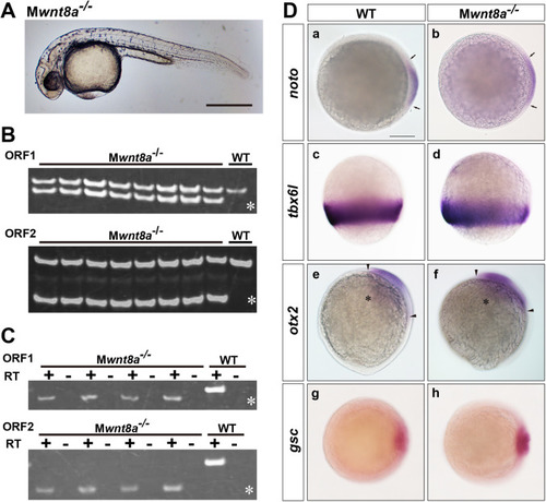
Maternal wnt8a mutants show no defects in dorsal-axis formation. (A) Maternal wnt8a mutant embryos (Mwnt8a-/-) at 30 dpf. Embryos were obtained by crossing WT male fish and female fish with wnt8aΔO1ΔO2/ΔO1ΔO2 germ cells. Lateral view with anterior to the left. The Mwnt8a-/- mutant embryos showed no apparent abnormality in dorsal axis formation (n=190). (B, C) Genotyping and RT-PCR of Mwnt8 mutant embryos. The TALEN target regions of ORF1 (upper panel) and ORF2 (lower panel) were amplified by PCR from the genome of a WT and eight Mwnt8a mutant embryos (B), and by RT-PCR from the RNA of a one-cell-stage WT and four Mwnt8a mutant embryos (C). The PCR products were separated on acrylamide gels. The positions of the PCR bands corresponding to the mutant DNAs are marked by asterisks. “+” and “-” indicate the presence and absence of reverse transcriptase (RT) in the cDNA synthesis. Note that all of the Mwnt8a mutant embryos had both WT and mutant alleles (n=8/8), and all four Mwnt8a mutants had only mutant ORF1 and ORF2 transcripts. (D) Expression of noto (shield stage, a, b), tbx6l (80% epiboly stage, c, d), otx2 (100% epiboly stage, e, f), and gsc (shield stage, g, h) in WT and Mwnt8a mutants. Mwnt8a mutants were obtained by crossing female fish with wnt8aΔO1ΔO2/ΔO1ΔO2 germ cells and WT (b, d, f) or wnt8aΔO1ΔO2/+ male (h) fish. Animal pole views with dorsal to the right (a, b, g, h). Lateral views with dorsal to the right (c-f). The expression of noto, tbx6l, otx2, and gsc was unaffected in the Mwnt8a mutants compared to WT embryos (noto, n=20; tbx6l, n=20; otx2, n=20; gsc, n=16). Scale bars: 500 µm in A; 200 µm in D. EXPRESSION / LABELING:
|
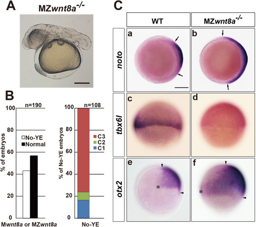
Maternal wnt8a supports the functions of zygotic wnt8a. (A) Phenotype of maternal-zygotic wnt8a mutant (MZwnt8a-/-) embryos at 30 hpf. Embryos were obtained by crossing wnt8aΔO1ΔO2/+ male fish and female fish with wnt8aΔO1ΔO2/ΔO1ΔO2 germ cells. Lateral view with anterior to the left. Approximately half of the embryos were predicted to be MZwnt8a-/- embryos, and the rest were Mwnt8a-/- embryos. The MZwnt8a-/- embryos were identified by genotyping. (B) Percentage of embryos showing the no-yolk extension phenotype (No-YE, left panel), and of No-YE embryos showing the antero-dorsalized phenotypes C1-3 (right panel, Fig. 2B). MZwnt8a showed more severe antero-dorsalized phenotypes than did zygotic (Z) wnt8a mutants (ΔO1ΔO2 in Fig. 2C). The proportions of C1-3 embryos were significantly different between the Zwnt8a and MZwnt8a mutants (chi-square test, P<0.001). (C) Expression of noto (shield-stage, a, b), tbx6l (60% epiboly, c, d), and otx2 (100% epiboly stage, e, f) in WT and MZwnt8a mutant embryos. Animal pole views with dorsal to the right (a, b). Lateral views with dorsal to the right (c-f). The lateral limits of noto expression are marked by arrows, the rostral and caudal limits of otx2 expression are marked by arrowheads, and the ventral limit of otx2 expression is marked by an asterisk. An expanded expression of noto, reduced expression of tbx6l, and expanded expression of otx2 were observed in approximately half of the MZ mutant embryos (noto, n=28/65; tbx6l, n=13/26; otx2, n=13/28), compared to WT embryos (normal expression of noto, n=30/30; tbx6l, n=20/20; otx2, n=26/26). Scale bars: 500 µm in A; 200 µm in C. EXPRESSION / LABELING:
|
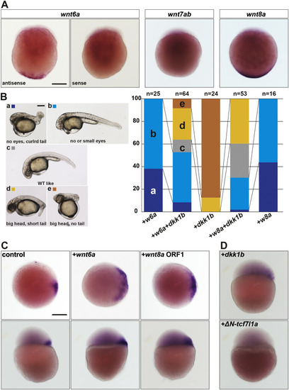
Wnt6a is another dorsal determinant candidate. (A) Maternal expression of wnt6a. Whole-mount in situ hybridization of wnt6a, wnt7ab, and wnt8 in one-cell-stage embryos. Staining with the sense probe for wnt6a was also performed as a negative control. wnt6a and wnt8a were detected in the vegetal pole (wnt6a antisense n=21/87, sense probe n=0/29; wnt8a n=18/19), whereas maternal wnt7ab was not detected (n=0/23). Lateral views. (B) Overexpression of wnt6a posteriorizes the embryos. One-cell-stage embryos were injected with the capped and polyadenylated RNA of wnt6a (5 pg), wnt8a (100 pg), dkk1b (50 pg), or a combination of wnt6a (5 pg) or wnt8a (100 pg), and dkk1b (50 pg). The phenotypes of the resultant embryos were observed at 30 hpf, and were categorized into five classes: no eyes, curled tail (a), no or small eyes (b), wild type-like (c), big head, short tail (d), and big head, no tail (e). Lateral views with anterior to the left. The proportion of embryos showing each phenotype is shown in the right panel. (C) Overexpression of wnt6a induces an expanded expression of dharma. The capped and polyadenylated RNA of wnt6a (1500 pg) or wnt8a (1500 pg) was injected into one-cell-stage embryos, and dharma expression was examined at the sphere stage. Animal pole views with dorsal to the right (upper panels); lateral views with dorsal to the right (lower panels). The dharma expression was restricted to the dorsal-most region (n=20/20), whereas it was expanded in embryos that received an injection of wnt6a RNA (n=12/42) or wnt8a RNA (n=27/64). (D) Overexpression of Dkk1b, a canonical Wnt pathway inhibitor, suppressed the dharma expression. Embryos were injected with dkk1b RNA (375 or 1500 pg) or ΔN-tcf7l1a RNA (900 pg, formerly called ΔN-tcf3a). Lateral views with dorsal to the right. The dharma expression at the sphere stage was reduced in the embryos that received an injection of dkk1b RNA (n=13/35 for 375 pg; n=8/20 for 1500 pg) or ΔN-tcf7l1a RNA (n=32/37). Scale bars: 200 µm in A; 500 µm in B; 200 µm in C. |

ZFIN is incorporating published figure images and captions as part of an ongoing project. Figures from some publications have not yet been curated, or are not available for display because of copyright restrictions. PHENOTYPE:
|
|
ZFIN is incorporating published figure images and captions as part of an ongoing project. Figures from some publications have not yet been curated, or are not available for display because of copyright restrictions. PHENOTYPE:
|
Reprinted from Developmental Biology, 434(1), Hino, H., Nakanishi, A., Seki, R., Aoki, T., Yamaha, E., Kawahara, A., Shimizu, T., Hibi, M., Roles of maternal wnt8a transcripts in axis formation in zebrafish, 96-107, Copyright (2017) with permission from Elsevier. Full text @ Dev. Biol.