
- Title
-
Phosphodiesterase Inhibitors Sildenafil and Vardenafil Reduce Zebrafish Rod Photoreceptor Outer Segment Shedding
- Authors
- Campbell, L.J., Jensen, A.M.
- Source
- Full text @ Invest. Ophthalmol. Vis. Sci.

Transgenic fluorescent marker is a tool for measuring rod outer segment renewal. (A) Schematic representation of a Tg(Xla.rho:EGFP); Tg(hsp70:HA-mCherryTM) rod photoreceptor with HA epitope– and transmembrane domain–tagged mCherry (red stripe) that has inserted into nascent rod discs and displaced toward the outer segment distal tip during the several days following a heat shock pulse. Growth distance (DG) is measured from the base of the outer segment to the mCherry stripe; shedding distance (DS) is measured from the mCherry stripe to the distal tip of the outer segment. (B, C) Representative z-projection images with DS measurements from (B) 8 dpf, 3 days post heat shock (dpHS), and (C) adult, 6 dpHS. Measurements are made in the three-dimensional z-stack. Photoreceptors express GFP (green) and are labeled with anti-Rhodopsin antibody (blue). |

Constant darkness reduces rod outer segment shedding. (A) Schematic timeline for examining rod outer segment (ROS) renewal in 8 dpf, 3 days post heat shock (dpHS) Tg(Xla.rho:EGFP); Tg(hsp70:HA-mCherryTM) fish after rearing in normal 14-hour light/10-hour dark cycle (LD) or constant dark (DD). (B) Representative images of the photoreceptor layer from LD- and DD-reared larvae immunolabeled with anti-GFP (green), anti-HA (red), and anti-Rhodopsin (blue) antibodies. Arrows in DD image indicate accumulation of phagosome-like bodies. Images are projections of a subset of z-sections totaling 11.09 μm. Scale bar: 10 μm. Quantification of (C) shedding distance (DS) and (D) growth distance (DG) in LD- and DD-reared larvae. Lower and upper hinges of box correspond to first and third quartiles; middle corresponds to median; whiskers extend 1.5 * interquartile range above and below the hinges; dots represent outliers. Graphs represent (C) n = 4 fish/treatment (96 LD, 157 DD outer segments) and (D) n = 4 fish/treatment (128 LD, 196 DD outer segments). (E) Schematic timeline for examining ROS renewal in adult Tg(Xla.rho:EGFP); Tg(hsp70:HA-mCherryTM) fish heat-shocked and reared 6 days in LD or DD. (F) Representative images of the photoreceptor layer from LD- and DD-reared adults immunolabeled with anti-GFP (green), anti-HA (red), and anti-Rhodopsin (blue) antibodies. Images are projections of a subset of z-sections totaling 5.97 μm. Scale bar: 10 μm. Quantification of (G) DS and (H) DG in LD- and DD-reared adults. Graphs represent (G) n = 3 fish/treatment (129 LD, 142 DD outer segments) and (H) n = 3 fish/treatment (187 LD, 190 DD outer segments). *95% CI of difference compared to control does not span zero. |
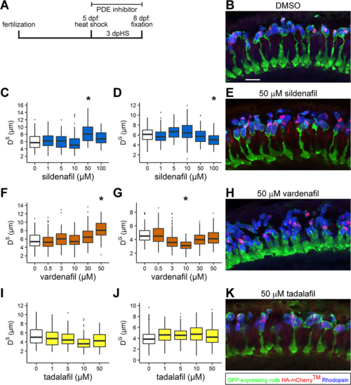
Sildenafil and vardenafil reduce larval rod outer segment shedding and vardenafil changes rod photoreceptor morphology. (A) Schematic timeline for examining rod outer segment renewal in 8 dpf, 3 days post heat shock (dpHS) Tg(Xla.rho:EGFP); Tg(hsp70:HA-mCherryTM); albb4/b4 fish after bathing in phosphodiesterase inhibitor. (B, E, H, K) Representative images of photoreceptor layers from 8 dpf, 3 dpHS larvae treated with (B) 0.05% DMSO, (E) 50 μM sildenafil, (H) 50 μM vardenafil, and (K) 50 μM tadalafil. Retinal sections were immunolabeled with anti-GFP (green), anti-HA (red), and anti-Rhodopsin (blue) antibodies. Images are projections of a subset of z-sections totaling 5.97 μm. Scale bar: 10 μm. Quantification of (C, F, I) shedding distance (DS) and (D, G, J) growth distance (DG). Lower and upper hinges of box correspond to first and third quartiles; middle corresponds to median; whiskers extend 1.5 * interquartile range above and below the hinges; dots represent outliers. Graphs represent (C, D) sildenafil n = 5 fish/treatment ([C] ≥63 and [D] ≥93 outer segments); (F, G) vardenafil n = 4 fish/treatment ([F]) ≥91 and [G] ≥122 outer segments); and (I, J) tadalafil n = 4 fish/treatment except for the 10 μM tadalafil sample with n = 3 fish ([I] ≥100 and [J] ≥190 outer segments). *95% CI of difference compared to control does not span zero. |

Sildenafil and vardenafil reduce adult rod outer segment shedding. (A) Schematic timeline for examining rod outer segment renewal in adult, 6 days post heat shock (dpHS) Tg(Xla.rho:EGFP); Tg(hsp70:HA-mCherryTM); albb4/b4 fish after bathing in phosphodiesterase inhibitor. (B) Representative images of photoreceptor layer from inhibitor-bathed adults immunolabeled with anti-GFP (green), anti-HA (red), and anti-Rhodopsin (blue) antibodies. Images are projections of a subset of z-sections totaling 5.97 μm. Scale bar: 10 μm. Quantification of (C) distance to shed (DS) and (D) growth distance (DG) in inhibitor-bathed adults. Lower and upper hinges of box correspond to first and third quartiles; middle corresponds to median; whiskers extend 1.5 * interquartile range above and below the hinges; dots represent outliers. Graphs represent (C) n = 3 fish/treatment (123 DMSO, 102 sildenafil, 133 vardenafil outer segments) and (D) n = 3 fish/treatment (176 DMSO, 192 sildenafil, 171 vardenafil outer segments). *95% CI of difference compared to control does not span zero. |

Rod outer segment shedding resumes following removal of sildenafil. (A) Schematic timeline for examining rod outer segment renewal in larval Tg(Xla.rho:EGFP); Tg(hsp70:HA-mCherryTM); albb4/b4 fish following heat shock at 5 dpf, sildenafil treatment for 3 days post heat shock (dpHS), and 1 to 3 days sildenafil removal (dWash). (B) Representative images of photoreceptor layers from 0 and 3 dWash sildenafil-treated larvae immunolabeled with anti-GFP (green), anti-HA (red), and anti-Rhodopsin (blue) antibodies. Images are projections of a subset of z-sections totaling 8.1 μm. Scale bar: 10 μm. Quantification of shedding distance (DS) following removal of (C) sildenafil for 0 to 3 days (dWash). Lower and upper hinges of box correspond to first and third quartiles; middle corresponds to median; whiskers extend 1.5 * interquartile range above and below the hinges; dots represent outliers. Graphs represent n = 5 retinas/condition (≥74 outer segments). *95% CI of difference compared to control does not span zero. |

Cone photoreceptors are sensitive to sildenafil and vardenafil. (A) Schematic timeline for examining green cone outer segment length in 8 dpf Tg(SWS1:mCherry); albb4/b4 larvae after bathing in phosphodiesterase inhibitor for 3 days (dTreat). (B) Representative images of photoreceptor layers from larvae treated with 0.05% DMSO, sildenafil, and vardenafil at indicated concentrations. Retinal sections were immunolabeled with green cone opsin antibody (green); ultraviolet cones express mCherry (red). Images are projections of a subset of z-sections totaling 7.33 μm. Scale bar: 10 μm. (C) Quantification of green cone outer segment full length (Dtotal). Lower and upper hinges of box correspond to first and third quartiles; middle corresponds to median; whiskers extend 1.5 * interquartile range above and below the hinges; dots represent outliers. Graph represents n = 5 fish/condition (DMSO = 162 outer segments, sildenafil = 169 outer segments, and vardenafil ≥ 175 outer segments). *95% CI of difference compared to control does not span zero. |