
- Title
-
The Cannabinoid Receptor Interacting Proteins 1 of zebrafish are not required for morphological development, viability or fertility
- Authors
- Fin, L., Bergamin, G., Steiner, R.A., Hughes, S.M.
- Source
- Full text @ Sci. Rep.

Accumulation of mRNAs from cnrip1a and cnrip1b genes. Whole mount in situ mRNA hybridisation of embryos at the indicated stages for antisense probes to cnrip1a (a) and cnrip1b (b). Lateral views (first two images in each panel) are anterior to top dorsal to left, except 24 hpf in which anterior is to left and dorsal to top. Dorsal views (remaining images) are anterior to left flatmounts (a) or wholemount (b), except panel a top right, which is a wholemount montage with anterior to top right. Sense control in inset in panel a top right is anterior to right dorsal to bottom. Quantitative evidence of reproducibility is given in Table S1. tel telencephalon, FB forebrain, MB midbrain, HB hindbrain, SC spinal cord, pf pectoral fin. Bars = 100 µm. EXPRESSION / LABELING:
|
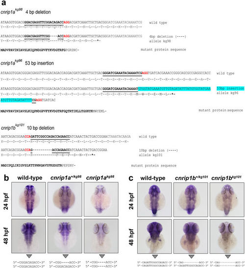
Mutagenesis of cnrip1a and cnrip1b. (a) Alignments of wild type with each of three mutant alleles showing the predicted expressed mutant polypeptide beneath. Bold underline indicates the gRNA target, red font the protospacer motif recognised by Cas9, hyphens deleted bases, blue highlight inserted bases and asterisks novel stop codons. In the mutant protein sequences, bold text indicates the residual wild type fragment and normal font the aberrant polypeptide tail. (b,c). To test for nonsense-mediated decay, about 50 siblings from in-crosses of cnrip1a +/kg98 (b) and cnrip1b +/kg101 (c) mutant carriers were subjected to in situ hybridisation for the cognate mRNA at the indicated stages, photographed, DNA extracted, PCR performed across the mutant locus and genotype confirmed by DNA sequencing as indicated beneath each panel. Quantitative evidence of reproducibility is given in Table S1. |

Maternal zygotic double cnrip1a kg98 ;cnrip1b kg101 mutant fish develop normally. (a) Schematic of crosses to generate a cnrip1 double mutant. (b) Darkfield image of 1 dpf double mutant indicating normal complex CNS folds (white arrowhead). (c) Differential interference contrast image of 1 dpf double mutant with arrowheads indicating normal eye (red), ear (purple), notochord (cyan) and haematopoetic tissue (black). (d) Dorsal, lateral and oblique (inset) views of 8 dpf larvae with arrowheads indicating pectoral fins (blue), jaw (turquoise), food traversing gut (green), swim bladder (orange) and xanthophores (yellow). Quantitative evidence of reproducibility is given in Table S1. All fish shown with anterior to left and dorsal up. Bars = 200 μm. |

ZFIN is incorporating published figure images and captions as part of an ongoing project. Figures from some publications have not yet been curated, or are not available for display because of copyright restrictions. PHENOTYPE:
|