- Title
-
SoxF factors induce Notch1 expression via direct transcriptional regulation during early arterial development
- Authors
- Chiang, I.K., Fritzsche, M., Pichol-Thievend, C., Neal, A., Holmes, K., Lagendijk, A., Overman, J., D'Angelo, D., Omini, A., Hermkens, D., Lesieur, E., Liu, K., Ratnayaka, I., Corada, M., Bou-Gharios, G., Carroll, J., Dejana, E., Schulte-Merker, S., Hogan, B., Beltrame, M., De Val, S., Francois, M.
- Source
- Full text @ Development
|
A SOX18-bound region within the notch1b locus represents a bona fide arterial-specific enhancer. (A) Part of the zebrafish notch1b locus from the UCSC ENCODE Genome Browser. The notch1b gene is in 3′ to 5′ orientation, H3K27ac peaks at 24 hpf are in purple, H3K4me1 peaks at 24 hpf are blue, SOX18Ragged ChIP-seq peaks are red, and the region encompassing the notch1b-15 enhancer is indicated by the purple horizontal bar. (B) The ZED notch1b-15:GFP transgene. Ins, insulator sequences; GATA2 prom, the silent GATA2 promoter; ZED control:RFP, the active cardiac actin enhancer/promoter construct fused to the RFP gene used as a positive control in the ZED vector. (C) The notch1b-15:GFP transgene directs arterial endothelial cell-specific expression in the zebrafish line tg(notch1b-15:GFP). Representative transgenic whole-mount embryos and transverse sections show reporter gene expression, as detected by in situ hybridisation (top row, blue) or GFP fluorescence (bottom rows, green) in arterial endothelial cells throughout embryonic development. nt, neural tube; nc, notochord; DA, dorsal aorta; PCV, posterior cardinal vein; aISV (arrows), arterial intersomitic vessel; DLAV, dorsal longitudinal anastomotic vessel. EXPRESSION / LABELING:
|
|
SoxF factors are required for notch1b-15 activity. (A) Confocal projection of stably transgenic WT tg(notch1b-15:GFP) (left) and tg(notch1b-15mutSOX-a/b:GFP) (right), where both zfSOX-a and zfSOX-b sites have been simultaneously mutated. Representative larvae from four independent founders are shown. Asterisks indicate the reduced GFP expression in the dorsal aorta. Arrowheads show the ectopic neuronal expression observed in tg(notch1b-15mutSOX-a/b:GFP). All panels show composite images from tile scan acquisition. (B,C) The intensity of GFP expression in the dorsal aorta and arterial intersomitic vessels of the stably transgenic tg(notch1b-15:GFP) and tg(notch1b-15mutSOX-a/b:GFP) at 2 dpf. Expression is normalised to GFP genomic copy number. Fish were pooled from three or four separate founders. Mean±s.e.m. tg(notch1b-15), n=41; tg(notch1b-15mutSOX-a/b:GFP), n=29. ****P<0.0001 (Mann–Whitney U-test). (D) Levels of GFP expression in tg(notch1b-15:GFP) zebrafish embryos after MO injection. Number of fish for each condition is indicated. (E) Representative examples of sox7/18 double-morphant tg(notch1b-15:GFP) zebrafish at 24 hpf as compared with uninjected controls. Double morphants (dMO) demonstrated reduced EGFP expression in the dorsal aorta and intersomitic vessels. DA, dorsal aorta; PCV, posterior cardinal vein; aISV, arterial intersomitic vessel. |
|
Loss of endogenous notch1b-15 compromises artery formation and reduces the endogenous notch1b transcript level. (A) The deleted region (dashed line) of notch1b-15 mutant allele uq1mf, which includes both the zfSOX-a and zfSOX-b sites (yellow). (B) F2 notch1b-15uq1mf/uq1mf has reduced notch1b expression in dorsal aorta and intersomitic vessels (white arrows) as compared with sibling WT and heterozygotes (black arrows) at 26-28 hpf. The number of embryos showing the illustrated phenotype among the total examined is indicated. (C) Quantitative PCR on FACS-sorted endothelial populations at 24-28 hpf, showing that F3 notch1b-15uq1mf/uq1mf fish have lower notch1b expression than WT fish. Expression is relative to kdrl and flt1. Mean±s.e.m. n=6 (uq1mf/uq1mf) and n=8 (WT) independent sorts, where each sort was pooled from 60-100 larvae;. *P<0.05, ***P<0.001 (t-test). (D) The treatment regime conducted to characterise the vascular phenotype of the uq1mf/+ cross. In treatment 1 (T1, red), notch1b MO was injected at the 1-2 cell stage. The developing vasculature of each embryo was then analysed blindly at 3 dpf. After scoring, genotypes were assigned to each larva. In treatment 2 (T2, blue), larvae from the uq1mf/+ cross were treated with or without DAPT (5 µM) from 15-16 hpf until 3 dpf. Vessels of these treated larvae were blindly scored prior to genotyping, as reported for T1. (E) At 3 dpf, notch1b-15uq1mf/uq1mf notch1b morphants frequently showed ectopic sprouting in between the intersomitic vessels (asterisks) as compared with sibling WT notch1b morphants. Mutants also show loss of arterial connections (red arrowheads) between the dorsal longitudinal anastomotic vessel (DLAV) and dorsal aorta (DA), as indicated by the loss of YFP expression in the tg(flt1:YFP) background. (F) (Top) Quantification of hypersprouting ISV number in individual notch1b morphants labelled by tg(flt1:YFP) at 3 dpf. Mean±s.e.m. Sibling WT (+/+), n=20; heterozygote (uq1mf/+), n=32; homozygous mutant (uq1mf/uq1mf), n=21. (Bottom) Quantification of YFP-positive intersomitic vessels that connect between DLAV and DA in individual notch1b morphants labelled by tg(flt1:YFP) at 3 dpf. Mean±s.e.m. Sibling WT, n=20; heterozygote, n=32; homozygous mutant, n=21. **P<0.005 (Mann–Whitney U-test). (G) Quantification of YFP-positive intersomitic vessels that connect between DLAV and DA in individual embryos treated with 5 μM DAPT at 3 dpf. Vessels are labelled by tg(flt1:YFP). Mean±s.e.m. Sibling WT, n=9; heterozygote, n=25; homozygous mutant, n=14. *P<0.05 (Mann–Whitney U-test). EXPRESSION / LABELING:
PHENOTYPE:
|
|
Mouse and zebrafish arterial Notch1 expression is dependent on SOX7/18 activity. (A) Transverse sections of whole-mount in situ hybridisation for Notch1 transcript on E8.5 mouse embryos shows a reduction of Notch1 expression (asterisks) in the bulbus cordis (bc) region of the primitive heart and vitelline artery (va) of Sox7/Sox18 double knockouts. (B) At 24 hpf notch1b expression is significantly downregulated in the dorsal aorta (asterisks) of sox7/sox18 double-morphant zebrafish, whereas its signal is unaffected in the neural tube. flt1 expression is comparable between controls and sox7/sox18 double morphants, indicating that the dorsal aorta is correctly formed. (C) notch1b was barely detectable in the dorsal aorta and ISVs of sox7/18 double-knockout zebrafish (asterisks), as compared with the WT, sox7 or sox18 heterozygotes (arrows). The number of embryos showing the illustrated phenotype among the total examined is indicated. DA, dorsal aorta; PCV, posterior cardinal vein. |
|
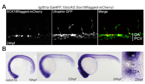
Expression of Sox18Ragged-mcherry and endogenous notch1b in developing zebrafish larvae. (A) The tg(fli1a:Gal4FF;10XUAS:Sox18Ragged-mcherry) line used to perform Sox18Ragged ChIP-Seq experiments. mCherry-Sox- 18Ragged fusion protein (red) could be observed in trunk blood vasculature (green). (B) notch1b mRNA is detected in arterial endothelial cells through-out embryonic development in a pattern similar to the notch1b-15:GFP transgene. nt = neural tube, nc = notochord, DA = dorsal aorta, PCV= posterior cardinal vein. EXPRESSION / LABELING:
|
|
Sox sites ablation compromises notch1b-15 enhancer activity. (A) A representative confocal projection of wild-type zebrafish F0 larvae injected with notch1b-15:GFP (top panel) and notch1b-15:mutSOX-a/b:GFP (bottom panel). Bright GFP expression could be observed in the intersomittic vessel and dorsal aorta of fish injected with the wild-type notch1b-15 construct, whereas expression of GFP in fish injected with the mutant construct is almost completely absent from the arterial vascular network (white arrows). (B) Graph indicating levels of GFP expression in F0 zebrafish transgenic for notch1b-15:GFP and notch1b-15mutSOX-a/b:GFP transgenes. All scored fish were also positive for the internal control cardiac actin:RFP (+RFP). Number of zebrafish examined for each conditions was indicated, expression was scored as no (-), weak or strong GFP . (C) Scatter plots representing the relationship between genomic GFP copy number and GFP intensity in endothelial lining of dorsal aorta (left panel) and intersomittic vessels (right panel) in stable tg(notch1b-15) and tg(notch1b-15mutSOX-a/b:GFP). Only larvae with GFP copy number <10 were included in the analysis for Fig. 7B-C. Scored tg(notch1b-15), n=46; tg(notch1b-15mutSOX-a/b:GFP), n=42; larvae were each pooled from 4 independent founders. |
|
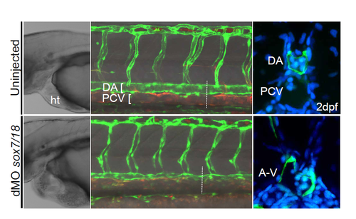
sox7/18 morpholino characterization. sox7/18 morpholino based knockdown of SoxF resulted in pericardial edema and fusion between the dorsal aorta and cardinal vein in zebrafish larvae. A-V = arterial venous shunts. PHENOTYPE:
|
|
Loss of endogenous notch1b-15 enhancer mildly phenocopies Notch1b loss-of-function. (A) A representative confocal projection of F2 homozygous zebrafish notch1b- 15uq1mf/uq1mf, derived from the F1 heterozygous notch1b-15uq1mf/+ in-crosses. No obvious phenotype was observed in the vasculature of F2 homozygous clutches. Quantification of YFP positive intersomittic vessels connecting between the dorsal longitudinal anastomotic vessel (DLAV) and dorsal aorta (DA) in individual F2 larvae labeled by tg(fltYFP;lyveDsred) at 3dpf (red arrowheads). Mean ± SEM; Scored sibling WT (+/+), n=19; Het (uq1mf/+), n=54; Hom (uq1mf/1mf), n=26; Mann-Whitney. (ns) No significant difference. (B) Representative confocal projections of F3 homozygous zebrafish notch1b-15uq1mf/uq1mf, derived from the F2 homozygous notch1b-15uq1mf/uq1mf in-crosses. While most F3 homozygous clutches lack phenotype (top left panel), a sub-population (20-30%) of the larvae showed either ectopic sprouting (asterisks) and/or loss of arterial connections (red arrowheads) between DLAV and DA, as indicated by the loss of YFP expression in tg(fltYFP;lyve1Dsred) background (bottom left panel). Quantification of YFP positive (top right panel) and hyper-sprouting (bottom right panel) intersomitic vessels in individual F3 larvae labeled by tg(fltYFP;lyveDsred) at 3dpf. Mean ± SEM; Scored WT (+/+), n=55; Hom (uq1mf/1mf), n=59; Mann-Whitney. (*) p<0.05, (***) p<0.001. |
|
Partial Notch inhibition results in the loss of arterial identity in notch1b-15 endogenous mutant fish. (A) At 3dpf, notch1b-15uq1mf/uq1mf mutant embryos treated with 5μM DAPT showed a significant reduction in the number of arterial connections (red arrowheads) between the dorsal longitudinal anastomotic vessel (DLAV) and dorsal aorta (DA), as indicated by the loss of YFP expression in the tg(fltYFP) background. (B) Quantification of YFP positive intersomitic vessels that connect between DLAV and DA in control (0μM DAPT) individual embryos at 3dpf. Vessels are labeled by tg(fltYFP). Mean ± SEM; scored sibling WT (+/+), n=3; Het (uq1mf/+), n=18; Hom (uq1mf/1mf), n=12; Mann-Whitney. (ns) No significant difference. |
|
Expression profiling of SOX7/18 mutant mice and zebrafish mophants. (A) Wholemount in situ hybridization assay showed a reduction of Notch1 expression in the primitive heart cavity and dorsal aorta (asterisks) in Sox7/Sox18 double knockouts at 8.5dpc (8-11ss) compared to the WT control and Sox7-/-; Sox18+/- control (black arrows). ht = heart, da = dorsal aorta. (B) Wholemount in situ hybirdization showed a comparable cdh5 expresison between control and sox7/18 double morphants, indicating that vascular structures were correctly formed in these morphants. |


