
- Title
-
Reprimo tissue-specific expression pattern is conserved between zebrafish and human
- Authors
- Figueroa, R.J., Carrasco-Avino, G., Wichmann, I.A., Lange, M., Owen, G.I., Siekmann, A.F., Corvalán, A.H., Opazo, J.C., Amigo, J.D.
- Source
- Full text @ PLoS One
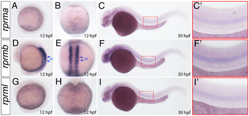
rprm expression patterns during early embryogenesis. (A-I) The expression patterns of rprma, rprmb and rprml were visualized by whole-mount in situ hybridization (WISH) during zebrafish embryonic development. Developmental stages are expressed as hours post-fertilization (hpf). (A, C, D, F, G, I) Lateral (anterior to the left) and (B, E, H) dorsal views are shown. Blue arrows in D-E indicate the expression in the somites (ss), while (C, F, I) the red inset magnifications indicate the expression in the notochord. EXPRESSION / LABELING:
|

RPRM expression patterns are conserved between zebrafish and human brain. (A-D) RPRM expression patterns were examined using whole-mount in situ hybridization in wild-type embryos at 28 and (E-H) 72 hours post-fertilization [hpf]. (A-D) Frontal views of the embryo heads. (E-H) Dorsal views of the embryo heads. (I-L) Retina cross-sections. (A-C) At 28 hpf rprma, rprmb and rprml transcripts are expressed in neuronal populations such as dorsal thalamus (DT), preoptic region (Po) and ventral thalamus, respectively (black arrows). (D) rprm3 is ubiquitously expressed throughout the brain. (E-G) At 72hpf rprma and rprml are expressed in the DT and the VT, respectively (white arrows), while rprmb is not expressed in those regions (asterisks). (H) At the same developmental stage, rprm3 mRNA is expressed in the Po and the optic chiasma (Oc). (I-L) Cross sections of the retina. (I, J, L) rprma, rprmb and rprm3 expression are restricted in the retina to the retinal ganglion cell layer (RGL, black arrow). (K) In contrast, rprml transcript expression is absent in the RGL (asterisk). (M, M’, N, N’) IHC staining for RPRM of white and grey matter sections from adult human samples (400x; inset magnifications 600x). (M-N) RPRM protein is expressed in the cytoplasm and axons of neurons (black arrowheads). (O-O’) RPRM protein is expressed in the nuclei of astrocytes. EXPRESSION / LABELING:
|

RPRM is expressed during zebrafish angiogenesis and in adult human vasculature. (A-C). Whole-mount in situ hybridization at 72hpf. Lateral view of the head vasculature. rprma/b and l are expressed in the mesencephalic vein (MsV), the dorsal longitudinal vein (DLV) and the primordial hindbrain channel (PHBC). (D-F) Lateral view of the trunk vessels. rprma/b and l are expressed at low levels in the dorsal aorta (DA, red bracket) the posterior cardinal vein (PCV, blue bracket) and the intersegmental vessels (ISV, red arrow). (D'-F') Inset magnifications (red brackets) and (D''-F'') transverse histological cross-sections of the posterior trunk region. (G-H) IHC staining for RPRM in tissue sections showing small blood vessels from adult human samples (400x; inset magnification 600x). (G, G') RPRM protein is expressed in the nuclei of endothelial cells (EC, black arrows) from a small artery. (H-H') RPRM protein is expressed in the nuclei of EC and vascular smooth muscle cells (vSMCs, black arrow) of a muscular artery. EXPRESSION / LABELING:
|
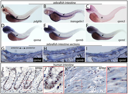
Expression of rprm genes during zebrafish gut development and human intestinal tissue. (A-F) Lateral views of whole-mount in situ hybridization (WISH) at 72hpf. (A-B) WISH for two well-known gut markers (A) platelet-derived growth factor receptor (pdgfrβ), (B) smooth muscle actin (sm22, also known as transgelin1), and the rprm genes (C) rprm3, (D) rprma (E) rprmb and (F) rprml. At 3dpf, rprma/-b and rprml are expressed in the embryonic intestinal tube. At the same developmental stage, there is a lack of expression of rprm3 in the intestinal tissue (C, asterisk). (G-I) Lateral cross sections of the zebrafish gut, anterior to the left. (J-J') RPRM IHC staining (400x; inset magnification 600x) showing nuclear positive expression in enterocytes at the base of the intestinal crypts. (K-K') RPRM IHC, (400x; inset magnification 600x) showing nuclear positive expression in smooth muscle cells from the muscularis propria of the small intestine. EXPRESSION / LABELING:
|

rprm expression during brain development in zebrafish embryos. rprm expression patterns were examined using whole-mount in situ hybridization in wild-type embryos at (A-D) 1 day post-fertilization [hpf]. (A-D) Lateral views. (A-C) At these developmental stages, rprma, rprmb and rprml transcripts are located in neuronal populations such as dorsal thalamus (DT), ventral thalamus (VT) and the cranial placode in the tigreminal ganglia (tg). (D) rprm3 is ubiquitously expressed through the brain. EXPRESSION / LABELING:
|

rprma and rprml are expressed in brain neurons in zebrafish larvae. (A-D) rprma and rprml expression was detected by whole-mount fluorescent in situ hybridization (FISH) at 72hours post-fertilization [hpf]. (A) Confocal cropping shows rprma expression in the anterior neurons of the telencephalon. (C-D) Confocal sectioning shows rprml expression in the posterior neurons within the forebrain (inset magnification). (A’D’) Transgenic Tg(fli1a:EGFP) is expressed in the endothelial cells within the major blood vessels of the head. |

rprma and rprmb are expressed during zebrafish angiogenesis. (A-B) Lateral views of whole-mount in situ hybridization at 48hpf. rprma/b are expressed in the mesencephalic vein (MsV), the dorsal longitudinal vein (DLV), the primordial hindbrain channel (PHBC), the primary head sinus (PHS), the nasal ciliary artery (NCA), the primary head sinus (PHS) and inner optic circle (IOC). Inset magnification shows that rprma/b are expressed dispersedly throughout the head vessels. (A'-B') Lateral views of the trunk vasculature, where rprma/b are expressed in hypochord (Hp, black arrow), the posterior cardinal vein (PCV, white bracket) and the intersegmental vessels (ISV, white arrows). EXPRESSION / LABELING:
|

FISH to localize rprm gene expression in zebrafish larvae. Top panels showing head or trunk views. (A-B) Vascular-specific transgenic Tg(fli1a:EGFP) marks endothelial cells and is revealed with the anti-GFP antibody. (C-F) FISH to detect rprma, rprml and pdgfrβ genes expression. pdgfrβ is used as mural cell marker. (G-H) merged confocal imaging. |