
- Title
-
Evolution of complexity in the zebrafish synapse proteome
- Authors
- Bayés, À., Collins, M.O., Reig-Viader, R., Gou, G., Goulding, D., Izquierdo, A., Choudhary, J.S., Emes, R.D., Grant, S.G.
- Source
- Full text @ Nat. Commun.

Transmission electron microscopy of zebrafish asymmetric synapses in four different brain areas. (a) Evolutionary tree of the vertebrate lineage with timescale in million years (my). The occurrence of the two WGD events common to all vertebrates (2R-WGD) and specific to teleosts (TSGD) are indicated by blue lines. (b) Schematic representation of the zebrafish brain with the four regions studied (CC, cerebellar corpus; O, olfactory bulb; OT, optic tectum; T, telencephalon) and of an excitatory synapse. Synaptosomes are formed by the axon terminal and the dendritic spine, which are separated from their corresponding neurons during tissue processing. The location of the PSD is also indicated. (c) Asymmetric synapse from the olfactory bulb. A red asterisk and a red arrow indicate the location of presynaptic vesicles and the PSD, respectively. Scale bar, 200 nm. (d) An asymmetric dendrodendritic synapse of the olfactory bulb (framed by a red dotted square) is shown. Asterisks indicate pre- and post-synaptic vesicles. The PSD is indicated by a red arrow. Scale bar, 500 nm. (e) Asymmetric synapses from the telencephalon. Red asterisks and arrows indicate the location of presynaptic vesicles and PSDs, respectively. The area corresponding to postsynaptic spine-like structures is filled with pink. Scale bar, 500 nm. (f) Asymmetric synapses from the optic tectum. Red asterisks and arrows indicate the location of presynaptic vesicles and the PSD, respectively. Red arrowheads indicate microtubule location within thin dendritic-like projection. The area of a thin dendritic-like projection, where synapses are formed, is filled with purple. Scale bar, 500 nm. (g) Flat (standard) asymmetric synapse from the cerebellar corpus. A red asterisk and a red arrow indicate the location of presynaptic vesicles and the PSD, respectively. Scale bar, 500 nm. (h) Asymmetric synapse from the medial part of the cerebellar corpus showing the extent at which the presynaptic element (highlighted in purple) surrounds the dendritic spine. Scale bar, 200 nm. (i) Micrograph displaying the morphology of most abundant asymmetric synapses from the cerebellar corpus. Red asterisks and arrows indicate the location of presynaptic vesicles and the PSD, respectively. Scale bar, 500 nm. |

Ultrastructure of synapses from Zebrafish olfactory bulb. (a) Schematic representation of the zebrafish brain. The black line indicates the position of the olfactory bulb. (b-i) PSDs found in different areas of the olfactory bulb. Arrowheads indicate PSDs from dendro-dendritic synapses. Scale bars b-c and e-i 500 nm; scale bar d 200 nm. |

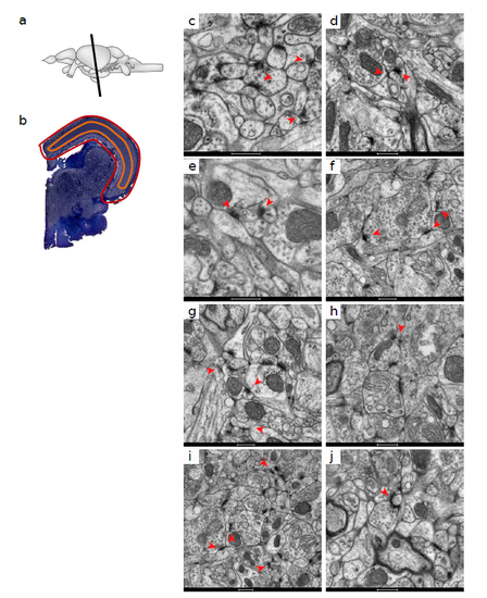
Ultrastructure of synapses from Zebrafish telencephalon. (a) Schematic representation of the zebrafish brain. The black line indicates the position of the telencephalon. (b) Coronal semi-thin section of the zebrafish telencephalon stained with toluidine blue. (c-j) PSDs found in different areas of the telencephalon. Scale bar c 1 μm; scale bars d-j 500 nm. |
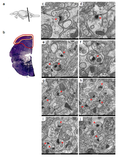
Ultrastructure of synapses from Zebrafish optic tectum. (a) Schematic representation of the zebrafish brain. The black line indicates the position of the midbrain. (b) Coronal semi-thin section of the zebrafish midbrain stained with toluidine blue. The red encircled area corresponds to the optic tectum. The orange line indicates the area where asymmetric synapses were identified. (c-j) PSDs found in different sections of the optic tectum. Arrowheads denote PSDs of postsynaptic elements showing either a small diameter or microtubules underneath. Scale bars c-j 500 nm. |
Ultrastructure of synapses from Zebrafish cerebellum. (a) Schematic representation of the zebrafish brain. The black line indicates the position of the hindbrain. (b) Coronal semi-thin section of the zebrafish hindbrain stained with toluidine blue. The red encircled area corresponds to the cerebellar corpus. The orange line indicates the area where asymmetric synapses were identified. (c-j) PSDs found in different sections of the cerebellum. Arrowheads indicate PSDs showing the curved morphology with the presynaptic element surrounding the postsynaptic spine. Scale bar c 200 nm; scale bars d-j 500 nm. |

Morphological characteristics of zebrafish PSDs a. Synapses from the cerebellar corpus with morphological characteristics similar to those observed in mammals and other zebrafish brain regions, including flat PSDs. Scale Bar 500nm. b. Synapses from the cerebellar corpus with presynaptic elements engulfing the postsynaptic terminal and highly curved PSDs. Scale Bar 500nm. c. Proportion of flat (light grey) and curved (dark grey) PSDs found at the cerebellar corpus of zebrafish brain. d. Representative images of the two types of PSD found amongst curved ones: type 1 (top image) and type 2 (bottom image). e. Representation of the three measures taken on cerebellar synapses: PSD arch length, PSD transversal length and spine longitudinal length. f. Histogram showing distribution of arch length for curved PSDs. g. PSD arch length differences between the three types of PSDs found at the cerebellar corpus (p < 0.05, Kruskal-Wallis test). Type 1 n = 24 PSDs; type 2 n = 40 PSDs; standard n = 10 PSDs. h. PSD transversal length differences between the two types of curved PSDs found at the cerebellar corpus. Type 1 n = 24 PSDs; type 2 n = 40 PSD. i. Spine longitudinal length differences between the two types of curved PSDs found at the cerebellar corpus. Type 1 n = 24 spines; type 2 n = 40 spines. j. Representation of the two measures taken on PSD from all brain regions: length and area. k. Differences in PSD length between brain regions (p < 0.05, Kruskal-Wallis test). Optic tectum n = 55 PSDs; Cerebellum type 1 n = 25 PSDs; Olfactory bulb n = 52 PSDs; Cerebellum standard n = 10 PSDs; Telencephalon n = 86 PSDs; Cerebellum type 2 n = 40 PSDs. l. Differences in PSD area between brain regions (p < 0.05, Kruskal-Wallis test). Olfactory bulb n = 53 PSDs; Optic tectum n = 53 PSDs; Cerebellum type 1 n = 26 PSDs; Cerebellum standard n = 10 PSDs; Telencephalon n = 87 PSDs; Cerebellum type 2 n = 37 PSDs. |