
- Title
-
Morphologic Changes of Zebrafish Melanophore after Intense Pulsed Light and Q-Switched Nd:YAG Laser Irradiation
- Authors
- Ryu, H.J., Lee, J.M., Jang, H.W., Park, H.C., Rhyu, I.J., Kim, I.H.
- Source
- Full text @ Ann. Dermatol.

Light microscopy in zebrafish before (A) and 1 week after (B) intense pulsed light (IPL) irradiation with 560 nm filter and 7 msec, and before (C) and 1 week after (D) IPL irradiation with 560 nm filter and 35 msec. Conventional IPL irradiation with a 7 msec pulse width induced melanophore breakage. However, the clearance of melanophore was not observed after irradiation with a 35 msec pulse width. |

Representative transmission electron microscope images of (A) control, (B) pulse-in-pulse mode intense pulsed light (IPL), (C) IPL irradiation with 560 nm filter and 7 msec, and (D) IPL irradiation with 560 nm filter and 35 msec. (B, C) Irradiation with pulse-in-pulse IPL and conventional IPL for 7 msec induced melanophore thermolysis (black arrowhead), with vacuolization (white arrowhead), central electron lucency (white arrow), and empty spaces (black arrow) evident due to melanophore destruction. Conventional IPL irradiation with 35 msec pulse width caused some melanophore changes but no vacuolization. Scale bar=100 µm. |
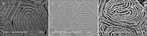
Representative scanning electron microscope images of (A) control, (B) Q-switched Nd:YAG (QSNY) laser irradiation, and (C) intense pulsed light (IPL) irradiation with 560 nm filter and 20 msec. Control zebrafish or zebrafish irradiated using a QSNY laser did not show microstructural changes. However, IPL-irradiated zebrafish showed finger-like fusion (arrows) in the protein structure of scales. |
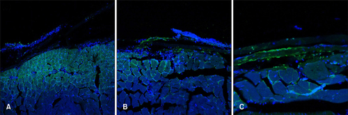
Representative confocal microscopic findings of (A) control, (B) pulse-in-pulsed mode intense pulsed light (IPL), and (C) conventional mode IPL irradiation with 560 nm filter and 7 msec. After conventional IPL irradiation, zebrafish showed a larger green-stained area on TUNEL staining than that after pulse-in-pulse mode IPL irradiation (×20). |

Normal adult zebrafish skin stained with H&E stain at ×100 magnification. Unlike the human skin, zebrafish have scales which are calcified plates originating in the dermis and covered by the mucous membrane. A horizontal view of fish skin reveals melanophores right below the scales. |