- Title
-
The inflammatory chemokine Cxcl18b exerts neutrophil-specific chemotaxis via the promiscuous chemokine receptor Cxcr2 in zebrafish
- Authors
- Torraca, V., Otto, N.A., Tavakoli-Tameh, A., Meijer, A.H.
- Source
- Full text @ Dev. Comp. Immunol.
|
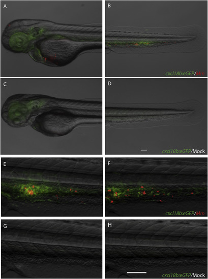
cxcl18b:eGFP is expressed by the tissue surrounding M. marinum infection sites. Injections of approximately 200 CFU of M. marinum M-mCrimson (Mm) via the caudal vein (A-B and E-F), but not mock injection (2% PVP in PBS, C-D and G-H) induced expression of the cxcl18b-eGFP transgene in the infected areas, especially localised around the sites of bacterial growth. Images were taken at 36 hpi. Scale bars: 100 μm. EXPRESSION / LABELING:
PHENOTYPE:
|
|
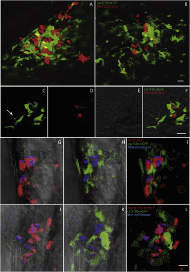
cxcl18b:eGFP is expressed by non-infected cells accumulated at the nascent granuloma. A-F. During the formation of granulomas, the cells that express high levels of cxcl18b-eGFP consist predominantly of uninfected cells that participate in the cell aggregates initiating the granuloma. Phenotypic inspection shows that some of these cells can emit long protrusions (arrow in C). G-L. Cxcl18b-expressing cells do not represent phagocytic cells since this marker does not overlap with established neutrophil and macrophage markers lyz (G–I) and mpeg1 (J–L). Images are representative of granulomas forming at 3 dpi (A–F) or 4 dpi (G–L). A-B are multiple z-stack maximum projections, C-L are split channels and overlay of an individual optical layer. In G-L outlines of green and red cells are marked with dashed lines of the corresponding colour. Scale bars: 10 μm. (For interpretation of the references to colour in this figure legend, the reader is referred to the web version of this article.) |


