
- Title
-
MicroRNA Profiling during Craniofacial Development: Potential Roles for Mir23b and Mir133b
- Authors
- Ding, H.L., Hooper, J.E., Batzel, P., Eames, B.F., Postlethwait, J.H., Artinger, K.B., Clouthier, D.E.
- Source
- Full text @ Front. Physiol.
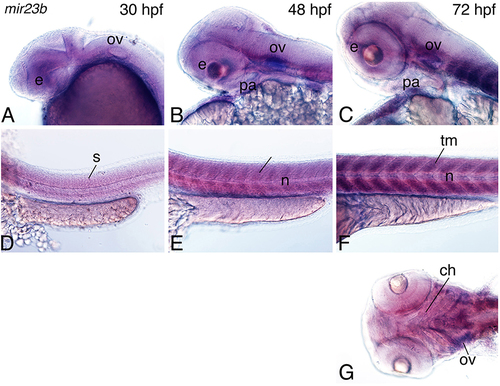
Figure 5. Expression of mir23b in zebrafish embryos. Whole-mount in situ hybridization analysis with a digoxigenin-labeled probe against the mir23b transcript at 30–72 hpf. (A–C) Expression is observed in the otic vesicle (ov; A), weakly in the eye (e), and pharyngeal arch (B,C), and palate (pl; C). (D–F) In the trunk, expression is observed in the trunk muscle the somites (s; E,D) at 30, 48 hpf, and trunk muscle (tm; F) at 72 hpf. (G) Ventral view of expression shows more detailed expression in the cranial muscle including the muscles supporting the craniofacial structures (arrows) and the eye. Anterior is to the left. n, notochord; ch, ceratohyal. EXPRESSION / LABELING:
|
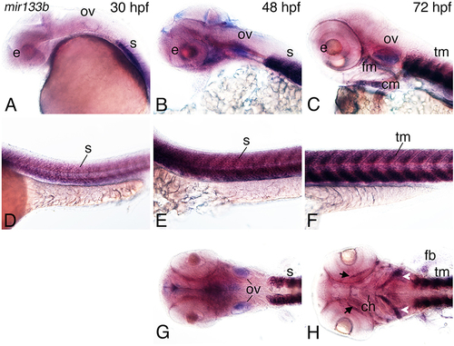
Figure 6. Expression of mir133b in zebrafish embryos. Whole-mount in situ hybridization analysis with a digoxigenin-labeled probe against mir133b at 30–72 hpf. (A–C) Expression is observed in the developing pharyngeal region, including at 48 hpf in the facial muscle (fm) and otic vessicle (B,G). (D–F) Strong expression of miR133b is observed in the somites (s; D) at 30 hpf, somites (E), and pectoral fin (pf) at 48 hpf (H) and facial muscle (fm; C), cardiac muscle (cm; C), and trunk muscle (tm; F) at 72 hpf, including the anterior mandibularis (arrows) and stemohyoideus (arrowheads) facial muscles muscles. Anterior is to the left. e, eye; tm, trunk muscle; n, notochord; ch, ceratohyal. EXPRESSION / LABELING:
|
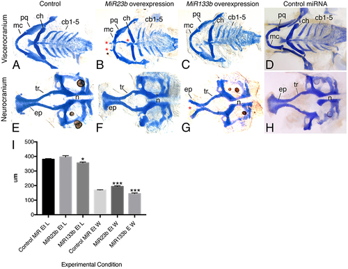
Figure 7. Injection of MiR133b and MiR23b RNA duplexes resulted in facial defects. Alcian blue stain of 6 dpf zebrafish larvae, flat mounted ventral views, anterior is to the left. (A,D,E,H) Control embryos, either uninjected or 33 μM control miR-injected, display wild type type craniofacial cartilage in both the viscerocranium and neurocranium. (B,F) Injection of 33 μM RNA duplex of MiR23b leds to a slight increase in the width of the ethmoid plate, likely resulting from shortening of the trabeculae (tr) (F), and production of ectopic cartilage (*; B) in the B) in the viscerocranium. (C,G) Injection of 6.25 μM RNA duplex of MiR133b resulted in hypoplastic cartilage structure and a cleft (*) in the ethmoid plate (ep; G,I). (I) Quantification of ethmoid plate length and width comparing standard control MiR (n = 10) injected with 33 μM compared to MiR23b (n = 9) and MiR133b (n = 9). Elements were measured and the mean and standard deviation for each are: Ethmoid plate length for control MiR, 380 ± 13.23 μm; MiR23b, 396 ± 27.13 μm; and MiR133b, 355 ± 19.27 μm; Ethmoid plate width for control MiR, 169.2 ± 4.92 μm; MiR23b, 193.9 ± 193.9 ± 14.09 μm; and MiR133b: 144.7 ± 14.7μm. Student's (Welsh's) T-test was used to compare to the standard control using GraphPad PRISM. *p < 0.02 and ***p < 0.0008. Anterior is to the left. cb, ceratobranchial; ch, ceratohyal; mc, Meckel's cartilage; n, notochord; pq, palatoquadrate. |

Whole mount ISH analysis miRNA expression in zebrafish embryos at 48 and 72 hpf. ISH analysis was performed using LNA probes. A, B. mir15a is expressed in the viscerocranium (vc) as well as the midbrain (mb) at 48 and 72 hpf. C, D. mir15b is expressed within the forming pharyngeal arch region at 48 hpf and expands to the neurocranium (n) and midbrain at 72 hpf. E, F. mir27b is expressed within the neurocranium, brain and heart (h) region at 48 and 72 hpf. G, H. mir30c expression is observed in the pharyngeal arches (pa) and brain at 48 hpf, and in the viscerocranium and trigeminal primordial (tg) at 72 hpf. I, J. mir130a is weakly expressed in the pharyngeal arch region at 48 hpf, and in the brain at 72 hpf. K, L. mir301a is expressed in the pharyngeal arches at 48 hpf and expands to the midbrain and trigeminal ganglia at 72 hpf. e, eye. EXPRESSION / LABELING:
|