- Title
-
Cavin4b/Murcb Is Required for Skeletal Muscle Development and Function in Zebrafish
- Authors
- Housley, M.P., Njaine, B., Ricciardi, F., Stone, O.A., Hölper, S., Krüger, M., Kostin, S., Stainier, D.Y.
- Source
- Full text @ PLoS Genet.
|
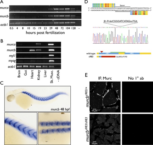
TALEN design and generation of a murcb mutant allele. A. RT-PCR analysis of mRNA expression of murca and murcb during zebrafish development. actb1 is shown as a loading control. B. RT-PCR analysis of mRNA expression of murca and murcb in adult zebrafish tissues. myl1 and myog are shown as controls for skeletal muscle contamination of other tissue samples. actb1 is shown as a loading control. C. Whole mount in situ hybridization of murcb mRNA in zebrafish embryos at 48 hpf. Top: lateral view. Bottom left: magnified lateral view. Bottom right: magnified dorsal view. D. TALEN construct for murcb mutagenesis. Sequencing results for the murcbs983 allele. Schematic of wild-type and predicted mutant proteins. The murcbs983 lesion leads to a premature stop codon after 83 missense amino acids starting at amino acid 28. Helical region (HR) domains are indicated in blue. The red arrowhead points to the TALEN target site. The red bar indicates the region of the mutant protein that is out of frame. The green bar indicates the antigen used to generate the antibody used in this work. E. Anti-Cavin4/Murc immunofluorescence (IF) micrographs of 10 µm transverse sections of skeletal muscle prepared from 10 wpf sibling and murcbs983/s983 fish. No 1° ab indicates control samples incubated only with secondary antibodies. PHENOTYPE:
|
|
Characterization of Cavin4b/Murcb deficient zebrafish. A. Representative lateral views of murcbs983/s983 and murcbs983/+ sibling zebrafish at 10 wpf. Fish in the top panels are females while those in the bottom panels are males. B. Dot plots of body mass and length of 10 wpf wt, murcbs983/+, and murcbs983/s983 zebrafish. Length measurements were made from the anterior point to the caudal fin/trunk boundary. Approximately equal numbers of male and female fish were used. The red line represents the mean. ***p<0.0005 C. H&E stain of 10 µm sections prepared from the trunk of 10 wpf murcbs983/+ and murcbs983/s983 zebrafish. Two examples of murcbs983/s983 fish of different sizes are shown. D. Phalloidin staining of 10 µm transverse sections prepared from the trunk of 10 wpf murcbs983/+ and murcbs983/s983 zebrafish. E. Trichrome staining of 10 µm transverse sections prepared from the trunk of 12 mpf murcbs983/+ and murcbs983/s983 zebrafish. F. Representative lateral views of murcbs983/+ and murcbs983/s983 zebrafish at 5 dpf. |
|
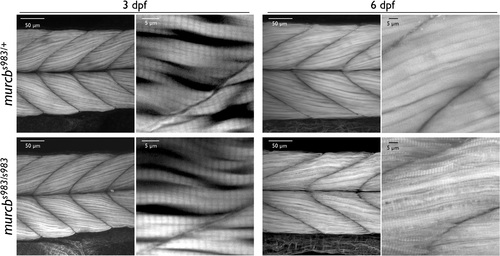
Filamentous actin analysis of Cavin4b/Murcb deficient zebrafish. Representative lateral views of confocal projections of phalloidin stained murcbs983/+ and murcbs983/s983 zebrafish at 3 dpf and 6 dpf. Skeletal muscle fibers are smaller and less consistent in size in murcb mutant animals compared to wild-type siblings. PHENOTYPE:
|
|
Swimming analysis of Cavin4b/Murcb deficient zebrafish. Dot plots of maximum swimming velocity and acceleration following the startle response of 60 hpf, 80 hpf, and 10 wpf murcbs983/+ and murcbs983/s983 zebrafish. The red line represents the mean. *p<0.05 ***p<0.0005 PHENOTYPE:
|
|
Ultra-structural analysis of Cavin4b/Murcb deficient skeletal muscle. A. Representative electron micrographs of murcbs983/+ and murcbs983/s983 zebrafish at 80 hpf. T-tubule triad structures are indicated with a box and enlargements of this region are shown on the left. B. Representative electron micrographs of murcbs983/+ and murcbs983/s983 zebrafish at 80 hpf. Caveloae are indicated by arrows. Enlargements from the micrographs are shown on the right. |
|
Caveolin expression and growth factor signaling in Cavin4b/Murcb deficient zebrafish. A. Representative confocal micrographs of Cav1 and Cav3 immunostaining of 10 µm transverse sections prepared from the trunk of 10 wpf murcbs983/+ and murcbs983/s983 zebrafish. B. Representative confocal micrographs of Cav1 whole mount immunostaining in murcbs983/+ and murcbs983/s983 zebrafish at 80 hpf. Mean relative fluorescence profiles from at least 6 larvae for each genotype. Pixel intensity was measured across a 3 µm segment covering a single striation and plotted relative to the background intensity between stria. SEM is represented by thin lines. C. Immunoblot of phospho-Thr202/Tyr204-Erk1/2 and total Erk1/2 from skeletal muscle prepared from 3 murcbs983/+ and 3 murcbs983/s983 10 wpf zebrafish. EXPRESSION / LABELING:
PHENOTYPE:
|
|
Murc conservation and expression in zebrafish. A. Protein alignment of zebrafish Murca and Murcb with human, mouse, rat, cow, and Xenopus orthologs. B. Zebrafish murc genes share a similar intron-exon structure with human MURC having two exons and a single intron. White boxes represent UTRs. C. Phylogenetic analysis of zebrafish Murca and Murcb. D. Whole mount in situ hybridization of murca mRNA in zebrafish embryos at 48 hpf. Top: lateral view. Bottom left: magnified lateral view. Bottom right: magnified dorsal view. EXPRESSION / LABELING:
|
|
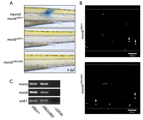
Analysis of Cavin4b/Murcb deficient zebrafish. A. Evans blue dye assay of membrane integrity. The top panel is an injured somite and is shown as a positive control. No significant changes were observed between mutants and heterozygous controls. B. Representative maximal projection confocal images from live whole mount acridine orange staining of murcbs983/+ and murcbs983/s983 zebrafish trunk at 80 hpf. Arrows point to DNA fragmentation. Dotted lines outline the larvae. No significant changes were observed between mutants and heterozygous controls. C. RT-PCR analysis of murca and murcb mRNA from murcbs983/+ and murcbs983/s983 larvae at 72 hpf. No significant changes were observed between mutants and heterozygous controls. |
|
Neuromuscular junction analysis of Cavin4b/Murcb deficient zebrafish. Representative confocal maximal projections of whole mount Synaptic Vesicle glycoprotein 2 (SV2, left) and α-bungarotoxin (α-BTX, right) staining of the trunk of 80 hpf murcbs983/+ and murcbs983/s983 zebrafish. SV2 immunofluorescence was used to visualize presynaptic structures; α-BTX staining was used to visualize postsynaptic structures. Synaptic punctae per somite were quantified using ImageJ (dotplot, bottom). PHENOTYPE:
|
|
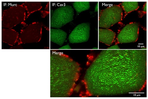
Immunofluorescence localization of Cav3 and Murc in adult skeletal muscle. Confocal micrographs of transverse sections prepared from 10 wpf murcbs983/+ zebrafish and stained with anti-Murc (red) and anti-Cav3 (green). Merged views are shown on the right and below. EXPRESSION / LABELING:
|


