
- Title
-
Individual Neuronal Subtypes Exhibit Diversity in CNS Myelination Mediated by Synaptic Vesicle Release
- Authors
- Koudelka, S., Voas, M.G., Almeida, R.G., Baraban, M., Soetaert, J., Meyer, M.P., Talbot, W.S., Lyons, D.A.
- Source
- Full text @ Curr. Biol.

GFP-Cntn1a as a Tool to Visualize Myelin Sheaths along Single Axons (A) Reticulospinal axon labeled with GFP-Cntn1a (top) in a Tg(sox10:mRFP) embryo, which labels oligodendrocytes and their myelin sheaths (middle). The myelin sheaths along the reticulospinal axon are localized to the gaps in GFP expression. Scale bar, 20 µm. (B) High-magnification views of areas outlined in (A) (top). Scale bar, 5 µm. GFP-Cntn1a fluorescent intensity profiles of the insets from (A) (bottom). (C) GFP-Cntn1a expression clustered at putative nodes of Ranvier (left) (arrows), as indicated by gaps in Tg(sox10:mRFP) expression (middle and right). Scale bar, 20 µm. (D) GFP-Cntn1A along a CoPA axon in a Tg(mbp:mCherry-CAAX) embryo at 4 dpf shows expression of the myelin reporter in the gap of GFP-Cntn1A localization. Scale bar, 5 µm. See also Figures S1 and S2 and Table S1. |

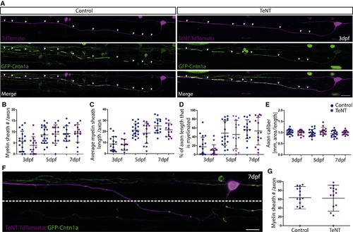
TeNT Expression in Reticulospinal Neurons Impairs Myelination along Individual Axons (A) Individual reticulospinal axons labeled with GFP-Cntn1a and TdTomato (left) and with GFP-Cntn1a and TeNT-Tdtomato (right) at 3 dpf, 5 dpf, and 7 dpf. Scale bar, 15 µm. (B-E) Quantification of myelin sheath number per axon per 425-µm imaging window (B), average length of myelin sheath per axon (C), percentage of axon length (per 425-µm imaging window) that is myelinated (D), and axon caliber (E) at 3 dpf, 5 dpf, and 7 dpf in control and TeNT-expressing reticulospinal neurons. All error bars indicate ± SD. See also Figure S3. |
TeNT Expression in CoPA Neurons Does Not Impair Myelination along Individual Axons (A) Individual CoPA axons labeled with GFP-cntn1a and TdTomato (left) and with GFP-Cntn1a and TeNT-TdTomato (right) at 3 dpf. Scale bar, 10 µm. (B-E) Quantification of myelin sheath number per axon (B), average length of myelin sheath per axon per 425-µm imaging window (C), percentage of axon length (per 425-µm imaging window) that is myelinated (D), and axon caliber (E) at 3 dpf, 5 dpf, and 7 dpf in control and TeNT expressing CoPA neurons. (F) Individual CoPA neuron and axon labeled with GFP-Cntn1a and TeNT-TdTomato. Dashed line indicates dorsoventral cutoff for axonal region analyzed when assessing region of CoPA axons in ventral spinal cord. Scale bar, 15 µm. (G) Percentage of axon length that is myelinated in the ventral spinal cord of control and TeNT expressing CoPA neurons at 7 dpf. All error bars indicate ± SD. See also Figure S3. |
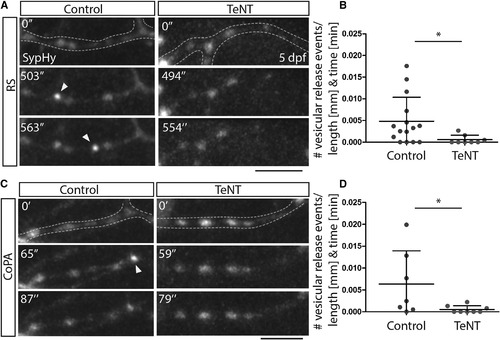
Tetanus Toxin Expression in Reticulospinal and CoPA Neurons Impairs Vesicular Release from Their Axons (A) Images from time-lapse movies of sypHy expression in reticulospinal axon collaterals in control (left) and TeNT-expressing (right) neurons at 5 dpf. Dashed lines outline the collateral. Arrowheads point to punctate increases in GFP expression indicative of vesicular release. Scale bar, 5 µm. (B) Quantitation indicates number of GFP events per collateral per micron per minute in control and TeNT-expressing reticulospinal neurons. (C) Images from time-lapse movies of sypHy expression in CoPA axon collaterals in control (left) and TeNT-expressing (right) neurons at 5 dpf. Dashed lines outline the collateral. Arrowheads point to punctate increases in GFP expression indicative of vesicular release. Scale bar, 5 µm. (D) Quantitation indicates number of GFP events per collateral per micron per minute in control and TeNT-expressing CoPA neurons. See also Movies S1, S2, S3, and S4. |
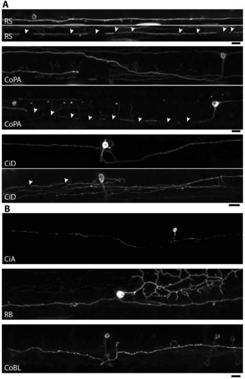
Overview of individual subtypes labelled with GFP-Cntn1a that have myelinated axons (A) and that do not have myelinated axons (B) over the stages examined. A shows an example of each neuronal subtype at stages prior to myelination and upon myelination. Arrows point to position of myelin sheaths. |

Left panels: Individual CoPA neuron lablelled with cytoplasmic GFP in a Tg(sox10:mRFP) animal, imaged with a 10x magnification objective lens at 3dpf (A) and 63x (B) and also at 5dpf (C). Arrows indicate myelin sheaths. Area imaged in B outlined by inset in A. Right panels: Individual CoPA neuron lablelled with GFP-Cntn1A in a Tg(sox10:mRFP) animal, imaged with a 10x magnification objective lens (D, top panel) 20x (D panels 2-4) and 63x (E). Arrows indicate myelin sheaths. Areas imaged in D outlined by insets in E. |

Individual reticulospinal axons (A) are of similar caliber when expressing control TdTomato (top) alone or TeNT-TdTomato (second from top) at 5 dpf. Scale bar: 15µm. Areas outlined by boxes in top and second panels are shown imaged by Airyscan in 3rd and 4th panels respectively. Scale bar: 5µm. Individual CoPA axons (B) are of similar caliber when expressing control TdTomato (top) alone or TeNT-TdTomato (second from top) at 5 dpf. Scale bar: 20µm. Areas outlined by boxes in top and second panels are shown imaged by Airyscan in 3rd and 4th panels respectively. Scale bar: 5µm. C and C′ show quantification of axonal caliber and reveal no difference between control and TeNT expressing reticulospinal neurons (C) or CoPA (C′). |