
- Title
-
Bmp signaling mediates endoderm pouch morphogenesis by regulating Fgf signaling in zebrafish
- Authors
- Lovely, C.B., Swartz, M.E., McCarthy, N., Norrie, J.L., Eberhart, J.K.
- Source
- Full text @ Development

The reception of Bmp signaling is temporally distinct between the endoderm and CNCCs. (A-K) Confocal images of endoderm (sox17:dsred) and Bmp-responsive cells (BRE:d2GFP) at the indicated stages of zebrafish development. (E-K) Orthogonal (yz-axis) views of A (E,F), B (G,H), C (I,J) and D (K). (L-S) Confocal images of CNCCs (sox10:mRFP) and Bmp-responsive cells (BRE:d2GFP). (P-S) Orthogonal views of L-O. Arrows indicate overlap between transgene expression; arrowheads denote lack of overlap. The endoderm is Bmp responsive by 14hpf (A-A′′) and this overlap persists to 26hpf (B-D′′), but only in the most posterior cells. The ventral CNCCs do not become Bmp responsive until after 18hpf (L-S). (A-D′′,L-O′′) Lateral views, anterior to the left. Scale bars: 50µm. EXPRESSION / LABELING:
|

Blocking Bmp signaling early disrupts endoderm morphology and craniofacial development. (A,B) Whole-mount images of viscerocranium at 5 days post-fertilization (dpf). Cartilage is blue and bone is red. Arrowheads point to missing cartilage elements and asterisks label the ceratobranchial cartilages. (A) No craniofacial defects are present following DMSO treatment. (B) Dorsomorphin (DM) treatment from 10-18hpf causes severe craniofacial defects. Ventral views, anterior to the left. Cartilages: Mc, Meckel′s; Ch, ceratohyal; Cb′s, ceratobranchial; Pq, palatoquadrate; HM, hyomandibular. (C,D) The percentage of viscerocranial defects in embryos treated with Dorsomorphin over multiple time windows. n values for all time points are listed in Table 1. (E,F) Confocal images of control and Dorsomorphin-treated sox17:DsRed embryos. (E′-F′′) Imaris surface renderings of E,F. E′′,F′′ are rotated 45° into the plane of view. (G,H) Confocal images of control or Dorsomorphin-treated sox10:mRFP embryos. Compared with controls (E-E′′,G), Dorsomorphin-treated embryos (F-F′′,H) had severe defects to both endoderm and CNCC morphology. Lateral views (except for E′′,F′′, see above), anterior to the left. P, pouch; PA, pharyngeal arch. Scale bars: 100µm in A; 50µm in E,G. |

Endoderm requires reception of Bmp signaling for proper development. (A-G) Whole-mount images (ventral view) of 5dpf viscerocranium. Asterisks label the ceratobranchial cartilages. (D′-E′′,G′) Confocal images (lateral view, anterior to the left) of 26hpf genetic chimeras labeling donor cells with either Alexa 568 dextran (D′,D′′), Alexa 488 dextran (E′,E′′) or the sox17:dsred transgene (G′). Arrowheads indicate endodermal pouch contribution, whereas arrows indicate endoderm failing to contribute to the pouches; insets show transgene expression with bright-field image. All embryos were heat shocked at 39°C for 1h at 10hpf. (A) Heat-shocked hs:dnBmpr1a-GFP embryos have severe craniofacial defects. (B) Control wild-type endoderm transplants do not disrupt craniofacial development. (D-D′′) Transplanting wild-type endoderm into a heat-shocked hs:dnBmpr1a-GFP embryo completely rescues craniofacial development (n=15/16). (E-E′′, arrows) Heat-shocked hs:dnBmpr1a-GFP donor cells are excluded from the pouches and fail to induce viscerocranial defects in wild-type embryos. (C) No rescue occurs when donor cells fail to populate the endoderm (n=6/6). (F) sox32 mutants lack endoderm and lose all of the viscerocranium. (G,G′) Heat-shocked hs:dnBmpr1a-GFP endoderm does not rescue sox32 mutants (n=6/6). Cartilages: Mc, Meckel′s; Ch, ceratohyal; Cb′s, ceratobranchial; Pq, palatoquadrate; HM, hyomandibular. Scale bars: 100µm in A-G; 50µm in D′-E′′,G′. PHENOTYPE:
|

Endoderm specification is maintained in Dorsomorphin-treated embryos. Images of 26hpf control or Dorsomorphin-treated embryos. (A,B) foxa1, (C,D) foxa2, (E,F) pdgfaa and (G,H) pdgfab are all expressed in Dorsomorphin-treated embryos. foxa1 expression is expanded, whereas foxa2 and pdgfab expression is reduced, and pdgfaa expression is normal. Pouch morphology is disrupted in all Dorsomorphin-treated embryos. (A-D) ventral views, (E-H) lateral views, anterior to the left. Scale bars: 50µm. |

Blocking Bmp signaling does not alter endodermal cell number. (A-F′,M) Confocal images of sox17:dsred transgenic embryos labeled for cell death at 18 and 26hpf. Similar, low levels of apoptosis are present across all groups (18hpf, f=0, P=1; 26hpf, f=0.3, P=0.6, one-way ANOVA, n=4 embryos). (G-L′,N) Confocal images of sox17:dsred transgenic embryos labeled for cell proliferation at 18 and 26hpf. No differences in the level of proliferating endoderm cells are apparent (18hpf, f=0.2, P=0.8; 26hpf, f=0.1, P=0.8, one-way ANOVA, n=4 embryos). Arrows indicate overlap between the transgene and marker expression. Lateral views, anterior to the left. (O) Total endoderm cell numbers were counted via FACS and no statistically significant differences between 26hpf Dorsomorphin-treated, untreated or DMSO-treated embryos were observed (f=1.8, P=0.2, one-way ANOVA, n=8 samples, 10 embryos per sample). Error bars indicate mean+s.e.m. Scale bar: 50µm. PHENOTYPE:
|
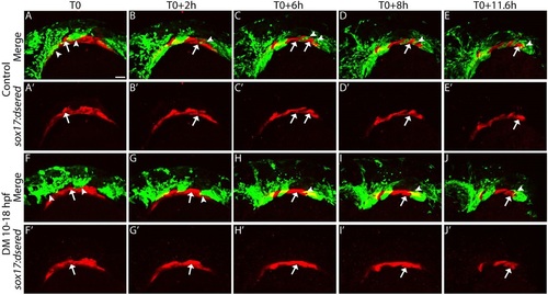
Bmp signaling regulates pouch morphogenesis. Still images from confocal time-lapse movies of sox17:dsred; sox10:EGFP double-transgenic embryos. (A,A′,F,F′) 18hpf embryos at the initiation of the movie (T0). (A-E′) Pouches (arrows) form in a sequential fashion to segregate the pharyngeal arches (arrowheads) in control embryos. (F-J′) In Dorsomorphin-treated embryos, pouch formation and arch segmentation fail. Lateral views, anterior to the left. Scale bar: 50µm. PHENOTYPE:
|

Fgf signaling activity requires Bmp. (A,B) Endodermal pouches express the Fgf signaling target etv4 at 30hpf in (A) control but not (B) Dorsomorphin-treated embryos. Lateral views, anterior to the left. (C-H) Expression of the Fgf receptors fgfr1b, fgfr3 and fgfr4 at 18hpf in control and Dorsomorphin-treated embryos. Only fgfr4 is expressed in the pharyngeal tissues and is reduced in Dorsomorphin-treated embryos (G,H, arrows). (C-F) Dorsal views, (G,H) ventral views, anterior to the left. Scale bars: 50µm. EXPRESSION / LABELING:
PHENOTYPE:
|
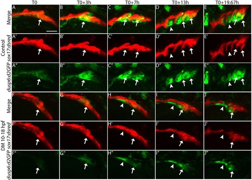
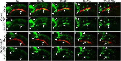
Blocking Bmp reduces the Fgf signaling response in the forming pouch. Still images from confocal time-lapse movies of sox17:dsred; dusp6:d2GFP (Fgf-responsive cells) double-transgenic embryos. (A-A′′,F-F′′) Embryos at 18hpf, the initiation of the movie (T0). Arrows indicate overlap between transgene expression, whereas arrowheads indicate lack of overlap. (F-J′′) Dorsomorphin-treated embryos show a reduced Fgf signaling response in the endoderm and pouches fail to form, in contrast to controls (A-E′′). Lateral views, anterior to the left. Scale bar: 50µm. PHENOTYPE:
|
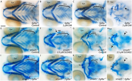
Bmp and Fgf signaling synergistically interact to achieve proper craniofacial development. Whole-mount images of 5dpf viscerocranium. (A-C) No gross craniofacial defects are present in wild-type or heterozygous controls (A), wild-type embryos treated with suboptimal doses of Dorsomorphin (B) or untreated fgf8a mutants (C). (D) Treating fgf8a mutants with suboptimal doses of Dorsomorphin causes severe craniofacial defects. (E,F) Similarly, wild-type or smad5 heterozygous embryos do not display any craniofacial defects (E), whereas these embryos treated with suboptimal doses of SU5402 (Pan-Fgf inhibitor, 10-18hpf) display minor hyomandibular defects (F, arrow). (G) Untreated smad5 mutants have characterized craniofacial defects (Swartz et al., 2011). (H) Suboptimal SU5402-treated smad5 mutants display severe craniofacial defects, including loss of ceratobranchial cartilages (arrowhead). (I-L) Severe craniofacial defects mirroring those of SU5402-treated smad5 mutants (see H) are present in smad5; fgf8a compound mutants (L) and not any other allelic combination (I-K). Ventral views, anterior to the left. Cartilages: Mc, Meckel′s; Ch, ceratohyal; Cb′s, ceratobranchial; Pq, palatoquadrate; HM, hyomandibular. Scale bar: 100µm. PHENOTYPE:
|

The reception of Bmp signaling in the CNCC′s but not the endoderm persist to at least 40 hpf. Confocal images of (A-C) sox17:dsred;BRE:d2GFP and (D-F) sox10:mRFP;BRE:d2GFP double transgenic embryos at (A&D) 32 hpf, (B&E) 36 hpf and (C&F) 40 hpf. Arrows show overlap between the transgene expression, arrowheads show where transgene expression does not overlap. The ventral CNCCs maintain a Bmp response out to at least 40 hpf (D-F), while the endoderm is not Bmp responsive during these time windows (A-C). Lateral views, anterior to the left. Scale bar = 50 µm. |
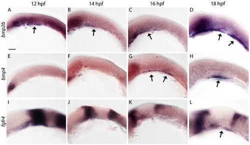
Bmp ligands are expressed in the developing pharyngeal arches prior to fgfr4. (A-H) Expression of the Bmp ligands, bmp2b (A-D) and bmp4 (E-H), and the Fgf receptor, fgfr4 (I-L), at 12, 14, 16 & 18 hpf. bmp2b is expressed in anteriorly developing pharyngeal tissues at 12, 14 and 16 hpf (A-C, arrows). Its expression is expanded at 18 hpf (D, arrows). Expression of bmp4 is broadly expressed at 16 hpf (G, arrows) which becomes more restricted by 18 hpf (H, arrow). Pharyngeal tissues expression fgfr4 only at 18 hpf (L, arrow), after expression of the Bmp ligands is observed. Lateral views, anterior to the left. Scale bar = 50 µm. |

Dorsomorphin treatment reduces Bmp signaling in the endoderm. (A-C) Confocal images of 18 hpf Dorsomorphin-treated sox17:dsred (endoderm) and BRE:d2GFP (Bmp response). Arrows show the reduced Bmp signaling response compared for Fig. 1 C-C′′. Lateral views, anterior to the left. Scale bars = 50 µm. |

Blocking Bmp signaling using a hs:dnBmpR1a-GFP transgenic line results in similar defects as Dorsomorphin treatment. (A&B) Whole-mount images of the viscerocranium of 5 dpf embryos labeled with Alcian Blue and Alizarin Red. (A) Control, heat-shocked embryos do not display any craniofacial defects. (B) The hs:dnBmpR1a-GFP embryos, heat-shocked at 10 hpf have severe craniofacial defects. (C&D) Images of heat-shocked sox17:dsred transgenic embryos. (C′-D′′) Imaris surface renderings of images in C&D, with C′′&D′′ being rotated 45° into the plane of view. (E&F) lateral views of sox10:mRFP. Embryos expressing hs:dnBmpR1a-GFP had defects to both endoderm and pharyngeal arch morphology. (A&B) ventral views, (C-F) lateral views, anterior to the left. Scale bar (A, B) = 100 µm, (C-F) = 50 µm, Mc = Meckel’s cartilage, Ch = ceratohyal cartilage, P = pouches PA = pharyngeal arches. |

Blocking Bmp signaling via hs:dnBmpR1a-GFP and loss of endoderm in sox32 mutants reduces Fgf signaling. (A&B) Expression of the Fgf signaling target etv4 in heat-shocked (A) control or (B) hs:dnBmpR1a-GFP embryos. (B) Expression of etv4 is reduced in heat-shocked hs:dnBmpR1a-GFP embryos compared to control embryos (A). (C&D) Expression of fgfr4 in wild-type (C) and sox32 mutant embryos (D). Expression of fgfr4 is reduced in embryos lacking endoderm (D compared to C). Lateral views (A&B), ventral views (C&D), anterior to the left. Scale bar = 50 µm. |
Blocking Bmp signaling reduces Fgf signaling response in the endoderm but not other Fgf responsive tissues. (A-J′) Expanded images from Figure 9. While endodermal expression of dusp6:d2GFP is reduced in Dorsomorphin-treated embryos (arrow), other expression domains, such as the trigeminal ganglion and mid-hindbrain boundary (arrowheads), appear normal. Lateral views, anterior to the left. Scale bar = 50 µm. |