
- Title
-
Intronic Flk1 Enhancer Directs Arterial-Specific Expression via RBPJ-Mediated Venous Repression
- Authors
- Becker, P.W., Sacilotto, N., Nornes, S., Neal, A., Thomas, M., Liu, K., Preece, C., Ratnayaka, I., Davies, B., Bou-Gharios, G., De Val, S.
- Source
- Full text @ Arterio., Thromb., and Vas. Bio.

The mouse Flk1in10 enhancer directs arterial-restricted expression in transgenic zebrafish. A, Schematic representation of the Flk1in10:GFP transgene. B, The mouse Flk1in10:GFP transgene directs endothelial cell-specific expression in transgenic zebrafish line tg(Flk1in10:GFP) at 24 h post fertilization (hpf). C-E, Tg(Flk1in10:GFP;kdrl:HRAS:mCherry) zebrafish expresses GFP in most endothelial cells at 30 hpf (C). At 48 hpf, GFP expression in the axial vessels becomes restricted to the dorsal aorta and to a subset of intersegmental vessels, corresponding to the developing intersegmental arteries (ISA; D; see also Movie I in the online-only Data Supplement). At 72 hpf, little dorsal aorta expression could be detected, while GFP expression is maintained in the ISA and dorsal longitudinal anastomotic vessel (E). F, Bar chart detailing GFP expression pattern in the ISA and intersegmental veins (ISVe) at 72 hpf. Represents a total of 18 embryos, ISA and ISVe identity established by using kdrl:HRAS-mCherry expression to determine whether each vessel connected to dorsal aorta or posterior cardinal vein. Any detectable level of GFP expression constituted positive, and Figure III in the online-only Data Supplement details analysis methods. bc indicates blood cells; da, dorsal aorta; dlav, dorsal longitudinal anastomotic vessel; dv, dorsal vein; GFP, green fluorescent protein; isv, intersegmental vessels; ISVe, intersegmental vein; and vv, ventral vein. |

The Flk1in10 enhancer requires ETS and GATA motifs for endothelial expression. A and B, Summary of reporter gene expression detected in (A) 48 hpf Tol2-mediated mosaic transient transgenic zebrafish embryos and (B) E12 transient transgenic mice. Asterisk indicates weak staining in head region only. C-F, Whole-mount (top) and transverse sections (bottom) of representative E12 X-gal-stained embryos transgenic for the Flk1in10mutGATA-b,c construct. Depicted sections correspond to the embryos shown. Number in bottom corner of whole-mount embryo denotes the number of embryos similar to that shown. Lines marked i and ii on C mark the approximate location of transverse sections. Arrowheads mark head vessels. G, Analysis of scrambled, 1.125 ng, 2.25 ng, and 3.375 ng gata2a morpholino (MO) in 48 h post fertilization (hpf) tg(Flk1in10:GFP) embryos. H, Analysis of scrambled, 2.25 ng and 3.375 ng gata2a MO in 26 hpf WT zebrafish embryos, using whole-mount kdr in situ hybridization. cv indicates cardinal vein; da, doral aorta; and GFP, green fluorescent protein. |

Rbpj is required for arterial restriction of the Flk1in10 enhancer. A and B, Summary of reporter gene expression detected in (A) 48 h post fertilization (hpf) Tol2-mediated mosaic transient transgenic zebrafish embryos and (B) embryonic day (E) 12 transient transgenic mice. C-F, Whole-mount (top) and transverse sections (bottom) of representative E12 X-gal-stained transient transgenic embryos expressing mouse Flk1in10 WT (C), mouse Flk1in10mRBPJ/SOX (D), mouse Flk1in10mRBPJ (E), and chicken Flk1in10 WT (F). Lines marked i and ii on C mark the approximate location of transverse sections in C-F. a indicates artery; cv, cardinal vein; da, dorsal aorta; and v, veins. G-I, Analysis of the effects of scrambled (G), 1.25 ng rbpj morpholino (MO) oligonucleotides (H), and Notch signaling inhibitor N-[N-3,5-difluorophenacetyl]-l-alanyl-S-phenylglycine methyl ester (DAPM) (I) in 72 h post fertilization (hpf) tg(Flk1in10:GFP) embryos. MO-mediated knockdown of rbpj results in expansion of Flk1in10:GFP expression into the caudal vein, the posterior cardinal vein, and intersegmental vein. da indicates dorsal aorta; GFP, green fluorescent protein; ISA, intersegmental artery; ISVe, intersegmental vein; and pcv, posterior cardinal vein. |
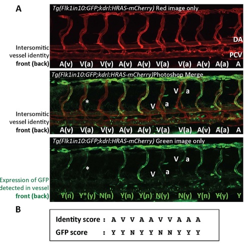
A. Detailed example of the analysis of arterial:venous identity of intersegmental sprouts used in Figure 2. Pictures denote single image of a representa+ve 72 hpf tg(Flk1in10:GFP;kdrl:HRASmCherry) embryo. Top panel denotes mCherry image only, in which the panvascular kdrl:HRASmCherry transgene is expressed in all intersegmental vessels as well as the dorsal aorta (DA) and posterior cardinal vein (PCV). Using this image we were able to assess the identity of each intersegmental vessel by whether it connects to the DA (arterial) or PCV (venous). The transparency of the embryo meant that we image two intersegmental vessels for each segment, one in the foreground denoted by UPPERCASE and the other in the background, denoted by bracketed lowercase. The middle image denotes both GFP and mCherry expression, and the bottom image just GFP expression. For each intersegmental vessel detected by kdrl:HRASmCherry expression, we scored whether GFP expression was detected (Y) or not (N). Vessels in foreground recorded as UPPERCASE, vessels in background as bracketed lowercase. * represents GFP in one cell, still recorded as GFP expression (Y) by our method. B. Creation of identity and GFP score used to create the bar chart in Figure 2. Records only foreground intersegmental identity and GFP expression. |

Loss of circulation does not prevent arterial restriction of Flk1in10:GFP expression. Analysis of the effect of 4ng tnnt2 MO in 28, 48 and 72 hpf tg(Flk1in10:GFP;kdrl:HRAS-mCherry) embryos. kdrl:HRASmCherry marks all vessels. ISA intersegmental artery, ISVe intersegmental vein. |

A. Analysis of scrambled, 2.25ng and 3.375 ng gata2a MO in 26 hpf WT zebrafish embryos, using whole-mount in situ hybridization with probes against arterial marker dll4, venous marker flt4 and kdrl. B. Analysis of 2.5ng gata1 MO in 36 hpf tg(Flk1in10:GFP) embryos. |