
- Title
-
Temporal and spatial requirements for Nodal-induced anterior mesendoderm and mesoderm in anterior neurulation
- Authors
- Gonsar, N., Coughlin, A., Clay-Wright, J.A., Borg, B.R., Kindt, L.M., Liang, J.O.
- Source
- Full text @ Genesis

Failure of neural tube closure in embryos treated with the Nodal inhibitor SB505124 for 20 min during mid blastula stages (3.8 and 4.0 hpf). Embryos treated with 75 µM SB505124 at all four stages of development have cyclopic eyes. The severity of the defects in mesendodermal/mesodermal derivatives, such as somites (brackets) decreases as the treatments move later in development. Embryos treated for 20 min at 3.8 and 4.0 hpf have open neural tubes (open NT), as evidenced by the two domains of pineal precursors (arrowheads). In contrast, embryos treated at 4.3 and 4.7 hpf have an oval shaped pineal anlage (arrowheads) indicating a closed anterior neural tube (closed NT). Top row: lateral views, animal pole to the top, scale bar: 250 µm. Second row: lateral views anterior to the left, scale bar: 50 µm. Third row: dorsal views anterior to the top. Scale bar: 50 µm. Refer to Table 1 for quantitative data. |
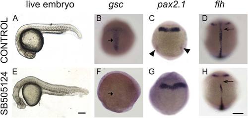
Exposure to Nodal inhibitor SB505124 at 3.8 hpf causes loss of mesendodermal and mesodermal derivatives. (A–D) Control WT embryos exposed to DMSO and (E–H) WT embryos exposed to 50 µM SB505124. (A) WT embryo at 1 dpf has a normal smooth and round head, a straight body axis, and complete somite formation, while (E) inhibitor-treated embryo displays cyclopia and abnormal morphology. (B) Normal gsc expression in the prechordal plate of a 9 hpf embryo (90% epiboly) displays a T-shaped expression domain (arrow) that (F) is absent in an inhibitor-treated embryo. (C) Normal pax2.1 expression is present along the midbrain–hindbrain border and in the posterior region in the pronephric mesoderm (arrowheads) in 10 hpf (tailbud stage) embryos. (G) In Nodal-deficient embryos, pax2.1 mRNA is present in the midbrain–hindbrain border (arrowheads) but absent in the pronephric mesoderm. (D) At 12 hpf (6 somite stage), flh is expressed in the developing notochord (arrow). (H) SB505124 treated embryos have partial reduction of notochord tissue. Scale bar: 250 µm. EXPRESSION / LABELING:
PHENOTYPE:
|
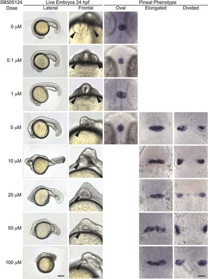
Open neural tube phenotype is fully penetrant upon treatment with SB505124 at concentrations of 10 µM and higher. All embryos were treated with the indicated concentration of SB505124 at 3.8 hpf (high oblong stage). First column: anterior to the left and dorsal to the top. Scale bar: 250 µm. Second column: frontal views. Scale bar: 100 µm. Arrowheads point to the eye. Third–fifth columns: Embryos were assayed for otx5 expression in the pineal. Dorsal views, anterior to the top. Scale bar: 50 µm. Refer to Table 2 for quantitative data. |

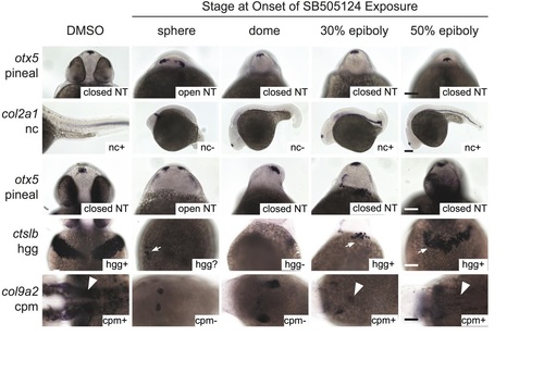
Initiating SB505124 treatment during mid blastula stages blocks neural tube closure. First column: developmental stage and images of live embryos at the onset of inhibitor exposure. Lateral views with animal pole to the top. Scale bar: 250 µm. Second column: lateral views of whole embryos with anterior to the left and dorsal to the top. Scale bar: 250 µm. Third–fifth column: Embryos were assayed for otx5 expression in the pineal gland. Dorsal views, anterior to the top. Scale bar: 50 µm. Refer to Table 3 for quantitative data. |
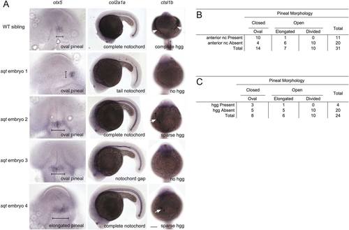
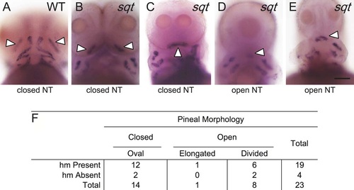
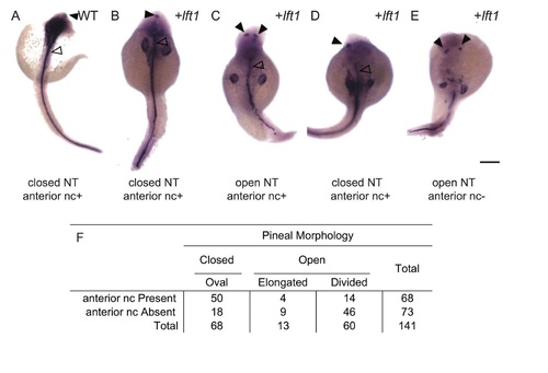
Positive correlation between neural tube closure and the presence of prechordal plate-derived hatching glands and axial mesoderm-derived anterior notochord. Embryos were raised to ~1 dpf and processed in tandem for otx5 in the pineal anlage, col2a1 in the notochord, and cstl1b in the hatching gland cells (hgg). (A) In WT embryos, the pineal organ is oval indicating a closed neural tube, the notochord spans the complete anterior–posterior extent of the trunk, and the hatching glands make a “necklace” around the anterior of the head. sqt embryos have a range of neural tube, notochord, and hgg defects as indicated. Note that, at this stage, col2a1 is expressed in three adjacent lines of cells, the floor plate (dorsal to the notochord), the hypochord (ventral to the notochord), and the notochord. The gaps in staining in sqt embryo 1 correspond to loss of all three tissues. The narrow region of staining in sqt embryo 3 just above the hind yolk corresponds to a place where the floor plate and hypochord cells persist, but the wider notochord cells are missing. The neural tube was scored as open if the pineal organ was at least twice as wide along its left–right axis (brackets) compared to its anterior–posterior axis. Images in the same row are different views of the same embryo. First column: dorsal views with anterior to the top. Second column: lateral views with anterior to the left and dorsal to the top. Third column: frontal views with anterior to the top. White arrows point to hatching glands. Scale bars: 100 µm. (B, C) Complete set of quantitative data. |
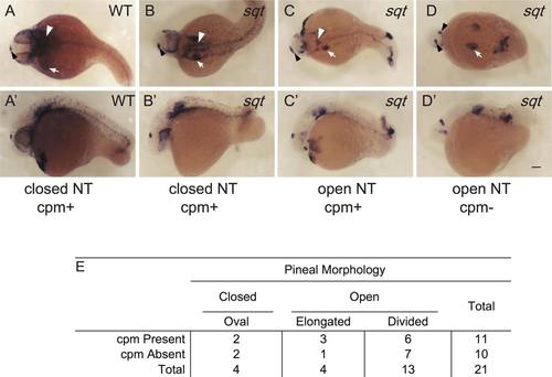
Correlation between cephalic paraxial mesoderm and neural tube closure. (A,A′) In WT embryos, col9a2 is expressed in the cpm (white arrowheads) and the ears (white arrow) and the otx5 expressing pineal anlage (black arrowheads) is oval shaped indicating a closed neural tube. (B–D′) sqt embryos with different combinations of cpm deficiencies and neural tube phenotypes. (A–D) Dorsal views with anterior to the left and (A′–D′) lateral views with anterior to the left and dorsal to the top. Images with the same letter are of the same embryo. (E) Complete set of quantitative data. Scale bar: 100 µm. |
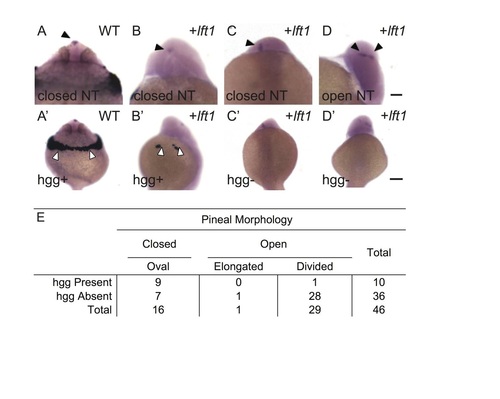
Correlation between presence of head muscles and neural tube closure in sqt mutants. (A–E) WT and sqt embryos were raised to 3 dpf and processed in tandem for smyhc2, in the head muscles (hm) and otx5, in the pineal anlage. The adductor mandibulae (am; white arrowheads) are the first muscles to develop in the head region and were used as an indicator for head muscle presence. (A) In WT embryos, am precursors are in two clusters just anterior to the yolk sac and other jaw muscles. The pineal organ is oval indicating a closed neural tube. (B–E) sqt embryos with an (B, C) oval pineal with the am present and (D, E) divided pineal with the am present. (F) Complete set of quantitative data. All images are frontal views with anterior to the top. Scale bar: 100 µm (A–E). EXPRESSION / LABELING:
PHENOTYPE:
|

Closed neural tubes and normal mesendoderm/mesoderm development in cyc and cas mutants. Embryos were coassayed for otx5 expression in the pineal precursors and a combination of the following mesendodermal/mesodermal markers: ctsl1b in the hatching glands, pax 2.1, col9a2 in cephalic paraxial mesoderm, and shh (for cas mutant) or col2a1 in the notochord (WT embryo and cyc mutant). Embryos in the first three rows are at ~26 somite stage and those in the last row are 30 hpf. Images in the same column and of the same age are different views of the same embryo. White arrowheads point to pineal, white arrows to hatching glands. Scale bars: 100 µm. EXPRESSION / LABELING:
|
[n/a] |
[n/a] |
[n/a] |

[n/a] |