- Title
-
Spatial and temporal expression of zebrafish glutathione peroxidase 4 a and b genes during early embryo development
- Authors
- Mendieta-Serrano, M.A., Schnabel-Peraza, D., Lomelí, H., Salas-Vidal, E.
- Source
- Full text @ Gene Expr. Patterns
|
Expression patterns of gpx4a and gpx4b during early zebrafish embryo development. A, RT-PCR expression analysis of gpx4a, gpx4b and 18s in zebrafish embryos at different developmental stages, 16- to 32-cell stage, sphere to dome, shield, 70%-epiboly, bud and 24 hpf. B, in situ hybridization analysis of gpx4a and gpx4b during early zebrafish embryo development. Upper inset, 36 hpf embryos were mounted in agarose and manually sliced to show the signal from the in situ localization of gpx4a is found in the periderm covering the yolk cell. Lower insets, highlight the gpx4b localization in the myotomes. Scale bar 1 mm for all stages and 250 µm for insets. |
|
GPx4 immunofluorescence localization patterns in whole zebrafish embryos from 1- to 8-cell stages as detected by laser confocal microscopy. 1-cell embryo (A, B, C and D). 4-cell stage embryo (E, F, G and H). 8-cell stage embryos (I, J, K and L). 8-cell stage control embryo in which the primary antibody was omitted but was incubated in the presence of secondary antibody (M, N and O). DNA, Hoechst-stained embryos (A, E. I and M). F-actin, phalloidin Alexa 488 stained embryos (B, F, J and N). GPx4, immunolocalization in whole embryos (C, G and K). Arrowheads indicate GPx4-positive signals in the yolk (C, G and K). Animal pole views are shown. Scale bar 200 µm. P, GPx4 antibody validation in zebrafish embryo samples at the shield stage. Gpx4a and b were down regulated by morpholino injection. A signal decrease was verified by western blotting, in which we compared immunoblot results with total protein samples from gpx4a or gpx4b morpholino injected embryos against standard control morpholino injected embryos. After immunoblotting for GPx4, the same membranes were stripped and immunoblotted against total ERK to confirm that the same amount of protein was loaded into the wells. EXPRESSION / LABELING:
PHENOTYPE:
|
|
GPx4 immunofluorescence localization patterns in 128- and 256-cell zebrafish embryos as detected by laser confocal microscopy. 128-cell embryo (A, B, C and D). 1, 2, 3, 4, 5, 6, 7 and 8 show amplifications of two different regions to highlight a cluster of cells that start showing GPx4 in the nuclei (6 and 8). 256-cell stage embryo (E, F, G and H). 9, 10, 11, 12, 13, 14, 15 and 16 are amplifications of two different regions that indicate the nuclear localization of GPx4. DNA, Hoechst-stained embryos (A and E). F-actin, phalloidin Alexa 488-stained embryos (B and F). GPx4, immunolocalization in whole embryos (C and G). Animal pole views are shown. A-H, scale bar 200 µm, insets 50 µm. EXPRESSION / LABELING:
|
|
GPx4 immunofluorescence localization patterns in zebrafish embryos at shield, 70%-epiboly and bud stages as detected by laser confocal microscopy. Shield stage embryos (A, B, C and D). 70%-epiboly (E, F, G and H). Bud stage embryos (I, J, K and L). Hoechst-stained embryos (A, E and I). F-actin, phalloidin Alexa 488-stained embryos (B, F and J). GPx4b immunolocalization (C, G and K). Left side views are shown. Dorsal side is to the right. Arrowheads indicate the epiboly migration front of the enveloping layer. Arrows indicate the position of the yolk nuclei epiboly migration front. Double arrowheads indicate tail bud position. *, evacuation zone. Scale bar 250 µm. |
|
GPx4 immunofluorescence localization patterns in zebrafish embryos at different somite stages as detected by laser confocal microscopy. 5-somite stage embryos (A, B, C and D). 14-somite stage embryos (E, F, G and H). 18-somite stage embryos embryos (I, J, K and L). Hoechst-stained embryos (A, E and I). F-actin, phalloidin Alexa 488-stained embryos (B, F and J). GPx4 immunolocalization (C, G and K). Left side views are shown. *, yolk cell which start to show GPx4a signal. Scale bar 250 µm. EXPRESSION / LABELING:
|
|
GPx4 immunofluorescence localization patterns in zebrafish embryos at 24 hpf as detected by laser confocal microscopy. Hoechst-stained embryos (A and D). F-actin, phalloidin Alexa 488-stained embryos (B and E). GPx4 immunolocalization (C and F). Embryos by 24 h of development (A, B and C) show GPx4b immunolocalization at the myotomes and at the longitudinal myosepta (C, arrow head). At slightly more advance stages (D, E and F) notice that the yolk cell extension (double asterisk) is of longer length and is narrower compared to younger embryos shown in A, B and C and show an increase in the immunlocalization signal apparently corresponding to GPx4a. The GPx4b immunolocalization is still found at the myotomes but is no longer found at the longitudinal myosepta (F). *, yolk cell. Scale bar 500 µm. EXPRESSION / LABELING:
|
|
GPx4b is localized in the slow muscle fibers 24 hpf zebrafish embryos myotomes. To identify in detail the GPx4b positive tissue in myotomes, different maximum intensity projections were generated from the confocal z optical slices. 1 (A to D), maximum intensity projections of full z stacks, representing approximately 25 µm. 2 (E to H), maximum intensity partial projections of slices located 5 µm depth shows that slow muscle fibers are located closer to the embryo surface, and the pattern of F-actin staining shows that slow muscle fibers are thicker and parallel to the main embryo axis (F). The GPx4b staining in slow muscle fibers is intense (G). 3 (I to L), deeper muscle fibers, at approximately 12 µm, show a different F-actin staining pattern, presenting thinner fibers (J) and a decreased GPx4 signal (K). Hoechst-stained embryos (A, E, and I). F-actin, phalloidin Alexa 488-stained embryos (B, F and J). GPx4b immunolocalization (C, G and K). Overlays (D, H and L), superimposition of F-actin (in red) and GPx4 (in green). M, orthogonal slice. Scale bar 50 µm. EXPRESSION / LABELING:
|
|
Expression patterns of gpx4a and gpx4b during early zebrafish embryo development. Embryos were analyzed at different developmental stages. Sense and antisense probes were included in the analysis to highlight the specificity of antisense in situ hybridizations. Scale bar 1 mm. |
|
Secondary antibody validation in zebrafish embryo samples at the different developmental stages. GPx4 antibody was omitted and unspecific secondary antibody binding to embryonic tissues was tested. We found that when embryos are processed with the same protocol but the primary antibody is omitted the secondary antibody give almost null signal when visualized with the same confocal imaging settings and during the same imaging sessions. Scale bar 250 µm. |
|
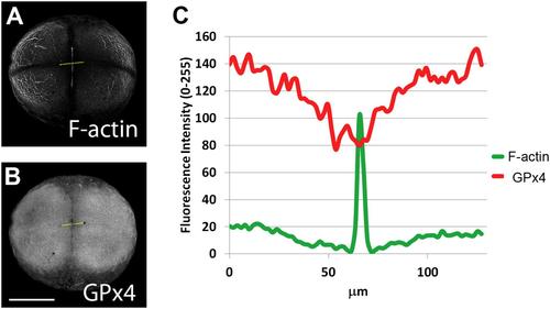
GPx4 protein is located at the blastomeres and is less abundant at the cleavage furrow (B), where important F-actin accumulation is found (A). Relative fluorescence intensity was determined by the gray scale pixel values along a line indicated in yellow (A and B) and plotted in the graph (C). Scale bar 250 µm. |
|
GPx4 localization patterns in zebrafish embryos at the 512-cell stage. Notice how the GPx4 nuclei localization is observed in all blastomeres at this stage (C). Overlay, superimposition of DNA (in blue), F-actin (in green) and GPx4 (in red). Animal pole views are shown. A-D, scale bar 200 µm, insets 50 µm. |

Unillustrated author statements PHENOTYPE:
|
Reprinted from Gene expression patterns : GEP, 19(1-2), Mendieta-Serrano, M.A., Schnabel-Peraza, D., Lomelí, H., Salas-Vidal, E., Spatial and temporal expression of zebrafish glutathione peroxidase 4 a and b genes during early embryo development, 98-107, Copyright (2015) with permission from Elsevier. Full text @ Gene Expr. Patterns


