
- Title
-
scaRNAs Regulate Splicing and Vertebrate Heart Development
- Authors
- Patil, P., Kibiryeva, N., Uechi, T., Marshall, J., O'Brien, J.E., Artman, M., Kenmochi, N., Bittel, D.C.
- Source
- Full text @ BBA Molecular Basis of Disease
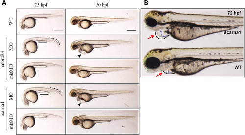
Heart deformities in MO-injected zebrafish. (A) Lateral views of wild-type and MO-injected embryos at 25 and 50 hpf. Both the snord94 and scarna1 morphants display little developmental delay with bent tail (black dotted curved line) and improperly formed yolk extension (black solid line) at 25 hpf. The pericardial edema (black triangle) and deformed yolk sac were more evident at 50 hpf. (B) Enlarged images of the heart region in wild-type and MO-injected embryos at 72 hpf. The conspicuous pericardial edema (black dotted circle) was observed in snoRNA MO-injected embryos. Gray outline is the atrium, blue outline is the ventricle. Scale bars: 200 µm. |
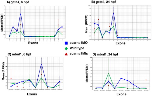
Representative examples of splice isoform variation (exon retention) after treating zebrafish embryos with anti-scarna1 morpholino. Each point on the horizontal axis represents an exon and the value represents the mean RPKM (reads per kilobase per million) for that exon. Gata4 and Mbnl1, 6 and 24 h post fertilization (hpf). |

Splice isoform changes after targeting scarna1. Treating zebrafish embryos with anti-scarna1 morpholino causes changes in exon retention of cardiac regulatory genes. 13 of 39 members of the Wnt family have significant changes in exon retention after treatment with antisense morpholino (assessed by RNA-Seq and qRT-PCR, values shown are from qRT-PCR data quantifying the variable exon). The misMO oligo is a negative control that differs from the primary morpholino by 5 nucleotides in the critical binding site. The gene name is shown on the horizontal axis and the numbers represent the start position of the exon. *Significantly different from the WT, p < 0.05. |

Cardiac abnormalities in MO-injected zebrafish. Lateral and ventral close-up images of the heart region in wild-type (WT) and embryos treated with antisense morpholino (MO) and mismatch morpholinos (misMOs) at 72 hpf. Heart is circled in the morphants treated with antisense morpholinos. Scale bars: 200 µm 31. PHENOTYPE:
|

ZFIN is incorporating published figure images and captions as part of an ongoing project. Figures from some publications have not yet been curated, or are not available for display because of copyright restrictions. PHENOTYPE:
|
|
ZFIN is incorporating published figure images and captions as part of an ongoing project. Figures from some publications have not yet been curated, or are not available for display because of copyright restrictions. PHENOTYPE:
|
Reprinted from Biochimica et biophysica acta. Molecular basis of disease, 1852(8), Patil, P., Kibiryeva, N., Uechi, T., Marshall, J., O'Brien, J.E., Artman, M., Kenmochi, N., Bittel, D.C., scaRNAs Regulate Splicing and Vertebrate Heart Development, 1619-29, Copyright (2015) with permission from Elsevier. Full text @ BBA Molecular Basis of Disease