|
jag1b gene expression is blocked in the gene-trapping jag1b mutant. (A) EGFP expression patterns in jag1b+/- embryos. EGFP is expressed in the otic vesicle (arrows). Scale bar: 100µm. (B) The zebrafish jag1b genomic locus. Primers used in D are indicated. (C) The mature wild-type (WT) jag1b transcript and the predicted mutant jag1b-EGFP fusion transcript. Primers used in E are indicated. (D) PCR genotyping confirms that the jag1b gene is disrupted in jag1b mutants (right). Wild type, heterozygotes and homozygotes show different GFP intensity (left). (E) RT-PCR analysis confirms the existence of the jag1b-EGFP fusion transcript in jag1b+/- and jag1b-/- embryos. (F,G) Real-time RT-PCR (F) and western blot (G) analyses indicate that jag1b mRNA and protein levels are decreased in jag1b mutant embryos. Error bars indicate s.e.m. of triplicated experiments. |
|
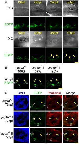
Anterior and posterior semicircular canal cristae are lost in jag1b-/- mutants. (A) High resolution EGFP expression patterns in the otic vesicle of jag1b+/- embryos. Images taken at 30hpf-48hpf are dorsolateral views. Other images are lateral views. (B,C) Confocal imaging (B) and phalloidin staining (C) of jag1b+/- and jag1b-/- embryos at 48hpf (B) and 72hpf (C). (B) Dorsolateral views. (C) Lateral views. Anterior towards the left and dorsal upwards. Scale bars: 50µm. AC, anterior crista; LC, lateral crista; PC, posterior crista. Arrows and arrowheads indicate sensory crista regions. EXPRESSION / LABELING:
PHENOTYPE:
|
|
Jag1b is required for survival of the posterior prosensory cells. (A) Confocal images of the otic vesicle of embryos at 24hpf. Lateral views. Scale bar: 50µm. (B) Whole-mount in situ hybridization of otic marker genes at 24hpf. Scale bar: 20µm. (C) Time-lapse live imaging of the developing posterior prosensory domain. The posterior sensory cells are gradually lost in jag1b-/- embryos. Scale bar: 10µm. (D) TUNEL staining of embryos at 34hpf and 40hpf. At 40hpf, apoptosis signals are detected only in the posterior prosensory domain of jag1b-/- embryos. Dorsolateral views. Scale bars: 40µm (left) and 20µm (right). Anterior towards the left and dorsal upwards. Boxed regions are enlarged on the right. |
|
fgf10a RNA levels are specifically reduced at the posterior prosensory domain in jag1b-/- embryos. (A-F) Whole-mount in situ hybridization of jag1b+/+, jag1b+/- and jag1b-/- embryos at 30 and 34hpf. Lateral views. Scale bar: 20µm. (G-H′′) Posterior cristae in jag1b+/- embryos treated with DMSO (G-G′′) or SU5402 (H-H′′) at 24-26hpf. Dorsolateral views. Scale bars: 50µm in G-H′; 10µm in G′′ and H′′. (I) TUNEL staining of jag1b+/- embryos treated with DMSO or SU5402. Apoptosis signals are detected in the posterior prosensory domain of the SU5402-treated jag1b+/- embryos at 42hpf. Dorsolateral views. Scale bars: 40µm (left) and 20µm (right). (J) Phalloidin staining of jag1b+/- embryos treated with DMSO or SU5402. Lateral views. Scale bar: 50µm. Anterior towards the left and dorsal upwards. Boxed regions are enlarged on the right. Arrows and arrowheads indicate posterior crista regions. EXPRESSION / LABELING:
|
|
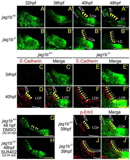
The anterior and lateral cristae are developed from a single anterior prosensory domain. (A-B′′′) Time-lapse imaging of the anterior pro-sensory domain development in jag1b+/- and jag1b-/- embryos. In jag1b-/- embryos, ACP and cells in the middle of the anterior prosensory domain undergo morphological change (B′′,B′′′, arrows). ACP, anterior crista primordia; LCP, lateral crista primordia; AC, anterior crista; LC, lateral crista. Arrows and arrowheads indicate cells undergoing morphological change. (C-F′) E-cadherin immunofluorescence staining in jag1b+/- and jag1b-/- embryos. At 40hpf, only cells in the middle of the anterior prosensory domain express E-cadherin in jag1b+/- embryos (D,D′, arrowheads). In jag1b-/- embryos, ACP and cells in the middle of the anterior prosensory domain express E-cadherin (F,F′, arrows). (G,H) Treatment with FGF inhibitor SU5402 at 32-34hpf blocks the separation of anterior and lateral cristae in jag1b+/- embryos. (I-J′) pErk3 immunofluorescent staining in jag1b+/- and jag1b-/- embryos. Dorsolateral views. Scale bars: 20µm. Anterior towards the left and dorsal upwards. Arrows and arrowheads indicate staining of E-Cadherin or p-Erk3. |
|
MAPK pathway inhibition blocks the separation of the anterior and lateral cristae. (A-L′) MAPKK inhibition represses expression of p-Erk3 and E-cadherin in both jag1b+/- and jag1b-/- embryos. Arrows and arrowheads indicate staining of E-Cadherin or p-Erk3. (M-M′′) In jag1b+/- embryos, treatment with the MAPKK inhibitors CI-1040 (M′) and PD0325901 (M′′) blocks separation of the anterior and lateral cristae. (N-N′′) In jag1b-/- embryos, the MAPKK inhibitors CI-1040 (N′) and PD0325901 (N′′) block the morphological change of cells in the anterior prosensory domain. Dorsolateral views. Scale bars: 20µm. Anterior towards the left and dorsal upwards. EXPRESSION / LABELING:
|
|
Inhibition of MAPK signaling generates a larger crista. (A-D) DIC confocal imaging of CI-1040-treated jag1b+/- embryos at 3dpf. Kinocilia are indicated by arrowheads. AC, anterior crista; LC, lateral crista. (E-H′′) Phalloidin staining of MAPKK inhibitor CI-1040-treated jag1b+/- embryos at 3dpf. Lateral views. Anterior towards the left and dorsal upwards. Scale bars: 40µm. Arrows and arrowheads indicate cells between AC and LC (E′′-G′′). (I) Whole-mount in situ hybridization of DMSO-and CI-1040-treated embryos. The probes used is msxC antisense. Arrowheads indicate cristae. (J) Total kinocilia number in the anterior and lateral cristae of DMSO-treated embryos and kinocilia number in the fused crista of CI-1040-treated embryos. Data are shown as mean±s.e.m. Statistical analysis was performed using a t-test. n indicates the number of inner ears for each experimental group. EXPRESSION / LABELING:
|
|
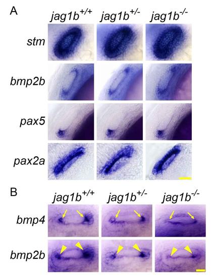
The expression of otic marker genes are similar among jag1b+/+, jag1b+/-, and jag1b-/- embryos. (A) Whole-mount in situ hybridization of otic marker genes at 24 hpf stage. The expression of otic maker genes: stm,bmp2b, pax2a, pax5 are similar among jag1b+/+, jag1b+/-, and jag1b-/- embryos. (B) Compared to wild type embryos, the expression of bmp4(arrows) and bmp2b(arrowheads) is downregulated in jag1b+/- embryos and dramatically reduced in jag1b-/- embryos.Scale bar: 20 µm. EXPRESSION / LABELING:
|
|
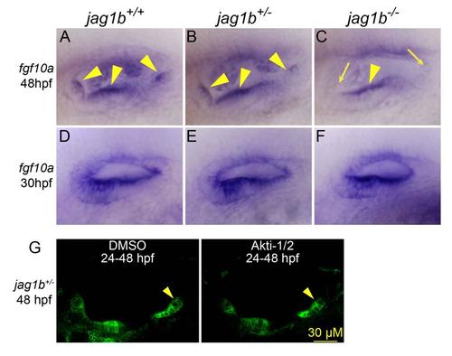
fgf10a is expressed in the sensory cristae. (A-C) Whole-mount in situ hybridization of fgf10a in jag1b+/+, jag1b+/-, and jag1b-/- embryos at 48 hpf stage. Probe: fgf10a anti-sense. fgf10a is expressed in the sensory cristae (arrow heads). In jag1b-/- embryos, the anterior and posterior cristae are lost. (D-E) Whole-mount in situ hybridization of fgf10a in jag1b+/+, jag1b+/-, and jag1b-/- embryos at 30 hpf stage. Probe: fgf10a anti-sense. In jag1b-/- embryos, fgf10a expression is not affected in the anterior prosensory domain. (G) Sensory cristae development is not affected by Akt inhibitor treatment. Lateral views with anterior to the left and dorsal up. |
|
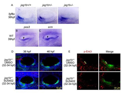
fgf8a is expressed in the anterior pro-sensory domain. (A) Whole-mount in situ hybridization, Probe: fgf8 anti-sense. fgf8 is expressed in the anterior prosensory domain at 36 hpf stage. jag1b+/+, jag1b+/-, and jag1b-/- embryos show similar fgf8a expression pattern. (B,C) Whole-mount in situ hybridization of pea3 (B) and erm (C) in WT embryos at 39 hpf stage. Probe: pea3 and erm anti-sense. pea3 and erm expression (arrowheads) is up-regulated in the middle region of the anterior prosensory domain. (D) TUNEL assay of DMSO and SU5402 treated embryos. (E) p-Erk3 immunostaining of DMSO and SU5402 treated embryos. Dorsolateral views with anterior to the left and dorsal up. EXPRESSION / LABELING:
|


