- Title
-
Endothelin signalling in iridophore development and stripe pattern formation of zebrafish
- Authors
- Krauss, J., Frohnhöfer, H.G., Walderich, B., Maischein, H.M., Weiler, C., Irion, U., Nüsslein-Volhard, C.
- Source
- Full text @ Biol. Open
|
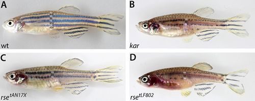
kar mutants display reductions in iridophores and melanophores similar to weak rse mutants. Wild-type (A), kar (B), weak rse (rse tAN17X) (C) and strong rse mutant (rsetLF802) (D) adult fish. The stripes (2D, 1D, 1V, 2V, 3V) and interstripes (X1D, X0, X1V, X2V) are indicated. Weak rse (C) and kar (B) display similar reductions in iridophores and melanophores as well as defects in the stripe pattern. PHENOTYPE:
|
|
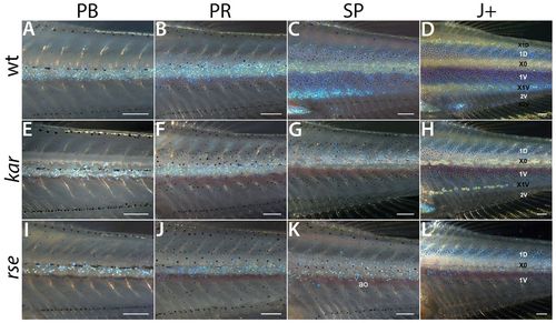
The kar phenotype arises during metamorphosis. Developmental series of body pigment pattern metamorphosis of wild-type (A–D), kar (E–H) and rsetLF802 mutant fish (I–L). Posterior trunk regions at the level of dorsal and anal fins are shown. The series of wild type and strong rse were published by Frohnhöfer et al. (Frohnhöfer et al., 2013). At stages PB and PR, kar mutants show only slight reductions in iridophore numbers (compare panels A,B to panels E,F), they still form a continuous sheet just ventral to the horizontal myoseptum. From PR to SP however, this reduction of iridophores becomes more apparent. They do not extend properly into dorsal and ventral interstripe regions (compare panel C and panel G), occasionally, remnants or patches develop (interstripe X1V in panel H). From stage SP onwards the number of melanophores is reduced compared to wild type (compare panels C,D to panels G,H). The early phase of pigment pattern development of strong rse mutants is similar to kar, with slightly stronger reductions in iridophores (I,J). In contrast to kar, however, iridophores do not form dense ridges in interstripe regions and rather are dispersed (K,L). ao: aorta. Scale bars: 250μm. PHENOTYPE:
|
|
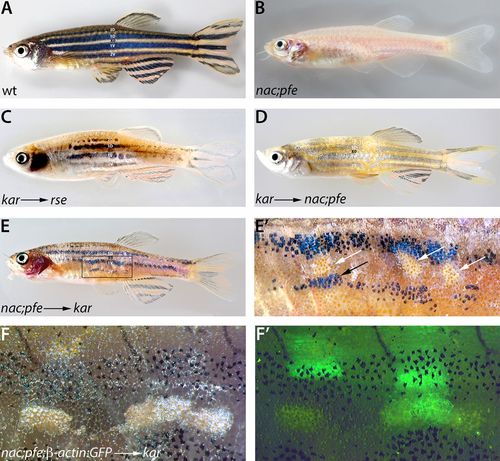
The kar gene product, Ece2, acts outside pigment cells to promote iridophore development. Wild-type (A) and nac;pfe mutant (B) adult fish for comparison. Transplantations of kar mutant cells into strong rse (C) or nac;pfe (D) mutant recipients result in fish with recovered stripe patterns. In chimeric fish generated by transplantation of nac;pfe donor cells into kar mutant recipient embryos (E,E2) occasionally, very small patches of dense iridophores develop (magnification in E2, white arrows). In the vicinity of these patches melanophores increase in number (black arrow). Labelling of the donors with Tg(β-actin:GFP) (F,F2) shows transplanted donor cells of various cell types next to the patches of dense iridophores. |
|
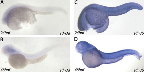
edn3b is expressed in the epidermis during early development. RNA in situ hybridizations for edn3a and edn3b at 24 hpf and 48 hpf in albino (alb) embryos. The expression of edn3a is not detectable above background (A,B). edn3b expression is detected in the epidermis during these stages (C,D). |
|
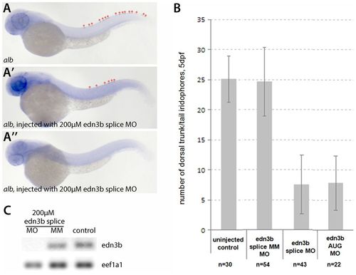
Morpholino-mediated knockdown of edn3b function in wild-type and alb embryos results in a strong reduction of iridophores. (A–A3) Morpholinos designed to interfere with splicing or translation of edn3b were injected into alb embryos, RNA in situ hybridization for pnp4a at 48 hpf shows a reduction of iridophore numbers in the dorsal stripes (marked with asterisks in panels A and A2). At 5 dpf morpholino injected larvae show strong reductions in iridophore numbers. Injection of a 5-bp-mismatch control morpholino had no effect (B). RT-PCR results to detect edn3b spliced message (C). Whereas the mismatch control morpholino showed no effect on the level of edn3b transcript, the PCR did not result in amplification of edn3b message after injection of the splice-interfering morpholino. PHENOTYPE:
|

