
- Title
-
Functional modelling of a novel mutation in BBS5
- Authors
- Al-Hamed, M.H., van Lennep, C., Hynes, A.M., Chrystal, P., Eley, L., Al-Fadhly, F., El Sayed, R., Simms, R.J., Meyer, B., Sayer, J.A.
- Source
- Full text @ Cilia
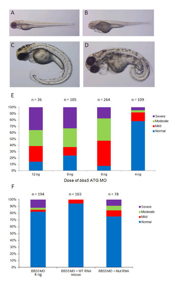
Phenotypic spectrum following bbs5 ATG MO injection and mRNA rescue. At 72 hpf following bbs5 MO injected (+/- mRNA rescue) zebrafish were phenotyped under light microscopy. Representative images are shown: (A) wildtype (WT) phenotype; (B) mildly affected morphant, showing a pericardial effusion and small eyes; (C) moderately affected morphant with a pericardial effusion, small eyes and a curly tail and (D) a severely affected morphant with a large pericardial effusion, small eyes and severe tail curvature. (E) Dose–response analysis of phenotypes following bbs5 ATG MO injection. Fish were classed phenotypically as normal if they were indistinguishable from uninjected fish; mildly affected if they had small eyes, mild pericardial effusion and mild/no tail defects; moderately affected if they had a large pericardial effusion and moderate tail defects, and severe if there was absent or very short malformed tail, widespread oedema, malformed eyes and minimal cardiac muscle contraction). Note a trend of increasing severity of phenotypes from 4 ng to 12 ng MO dose. The number of embryos (n) injected for each dose are shown. (F) Phenotypes of bbs5 ATG MO injected fish compared to co-injected WT BBS5 mRNA and mutant (Mut) BBS5 mRNA. PHENOTYPE:
|
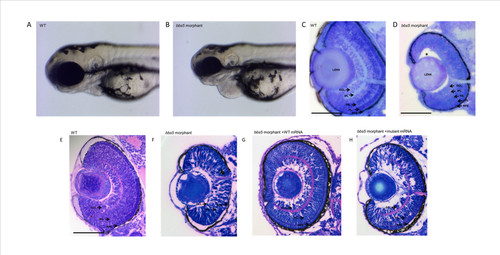
Microphthalmia and retinal layering defect in bbs5 morphants and phenotypic rescue with mRNA co-injection. Light microscopy of 72 hpf embryos show a reduction in eye size in between (A) wildtype (WT) and (B)bbs5 morphants. Retinal sections demonstrating (C) WT retina with normal, well demarcated retinal layering compared to (D) morphant retina displaying partial loss of retinal layering, loss of photoreceptor layer and separation of lens from retina (*). In mRNA rescue experiments, WT mRNA or mutant mRNA was co-injected with bbs5 MO. (E) Retinal layers are preserved in WT and (F) disrupted in bbs5 morphants. (G) WT mRNA is able to rescue the retinal phenotype and eye size whilst in the (H) mutant mRNA phenotype retinal layers remain disrupted and the eye size small. Scale bar 100 um. GCL, ganglion cell layer; INL, inner nuclear layer; IPL, inner plexiform layer; PL, photoreceptor layer, RPE, retinal pigment epithelia. PHENOTYPE:
|
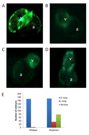
bbs5 morphants display cardiac anomalies including situs inversus. Wildtype (WT) and morphant cmlc2:GFP embryos were imaged at 48 hpf. (A) WT heart showing normal looping (D-looping) with the atrium (a) on the left hand side of the embryo and the ventricle (v) on the right. (B) Morphant heart with preserved D-looping. (C) Morphant heart with looping in the opposite direction (L-looping) resulting in the atrium on the right of the embryo and the ventricle on the left. (D) Morphant heart, which shows a failure of looping, resulting in a heart with neither the atrium nor ventricle leaving the midline. (E) 98% of WT embryos displayed D-looping whilst just 61% of morphant embryos exhibited D-looping, 12% exhibited L-looping, and 27% of morphant hearts were unlooped (P < 0.001). |
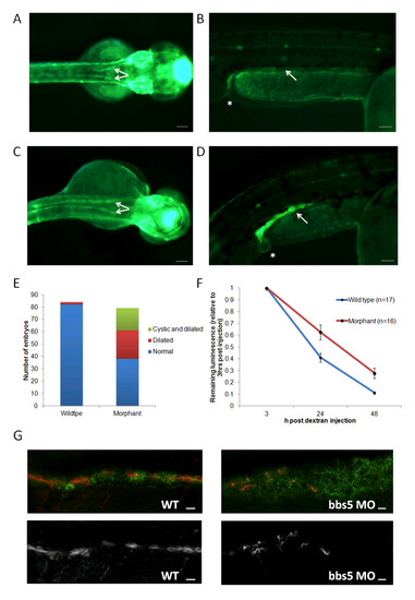
Characterisation of pronephros structure and function in bbs5 morphants. (A-D) Superior and lateral views of a cldnb:lynGFP embryo displaying (A,B) normal pronephric ducts (arrowed) and cloaca (*) in wildtype (WT) zebrafish. (C,D)bbs5 morphant embryos displayed dilated and tortuous pronephric ducts (identified by GFP fluorescence) and a (D) cloacal cystic dilatation. Scale bars are 100 μm. (E) Quantification of pronephros abnormalities in WT and bbs5 morphant embryos. (F) Estimation of GFR in zebrafish embryos was performed by measuring change in cardiac luminosity of both WT and morphant embryos after cardiac sinus fluorescent dextran injection. Mean luminescence +/- SEM (arbitrary units) is plotted versus time, up to 48 hours post injection. Morphant embryos retained significantly more fluorescent dextran at 24 hours (P = 0.005) and at 48 hours (P = 0.002). (G) At 72 hpf, compared to WT control, bbs5 morphant embryos revealed disrupted and fewer numbers of cilia in the dilated pronephros. Scale bar 10 μm). |
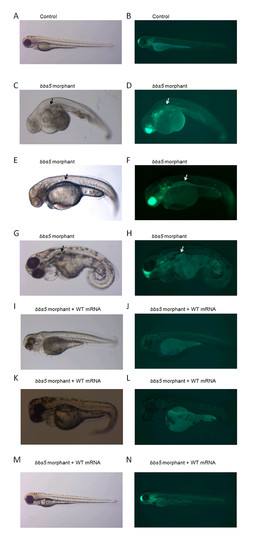
Light and fluorescence microscopy of renal cysts in bbs5 morphants and rescue with WT bbs5 mRNA. Left panels show bright-field images of 72 hpf embryos and right panels show immunofluorescence images, using claudin-Lyn-GFP embryos which express GFP throughout the pronephros (as well as forebrain and ear). (A,B) Uninjected fish (Control). (C-H) Morphological defects are seen in bb5 morphant embryos. bbs5 morphant embryos show pronephros dilatation and cyst formation which is subtle on light microscopy (black arrows) but more easily identified under fluorescence microscopy (white arrows). (I-N) Morphant phenotypes of tail abnormalities, pronephric duct dilatation /cysts are (I-L) partially and (M,N) fully rescued by co-injection with WT bbs5 mRNA. |