
- Title
-
The zebrafish amyloid precursor protein-b is required for motor neuron guidance and synapse formation
- Authors
- Abramsson, A., Kettunen, P., Banote, R.K., Lott, E., Li, M., Arner, A., and Zetterberg, H.
- Source
- Full text @ Dev. Biol.
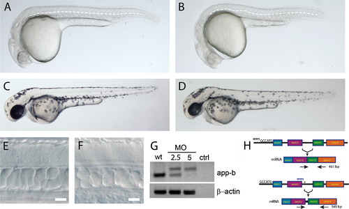
Partial knock-down of appb. Injection of 2.5 ng morpholino (MO) designed against appb results in an undulating notochord (B, dashed line) not present in animals injected with control MO at 24 hpf (A). Otherwise, the gross morphology of embryos injected with control (C) or appb morpholino (D) is comparable at 48 hpf. Cells within the notochord of appb morphants (F) are less vacuolated compared to controls (E). RT-PCR amplification of exon 1 and 4 (G,H) results in a larger fragment in embryos injected with appb morpholino than control morpholino (G). At 2.5 ng approximately 50–60% of the wildtype transcript was present, whereas it was absent at 5 ng morpholino. (H) Schematic illustration of wildtype and trslMO splicing (upper) and the effect of the appb morpholino (blue comb) on splicing. Blocking the splicing of intron 2 activates an alternative splice site (N) within intron 2 resulting in a premature stop in exon 3 (H). Scale bar: 20 μm. |
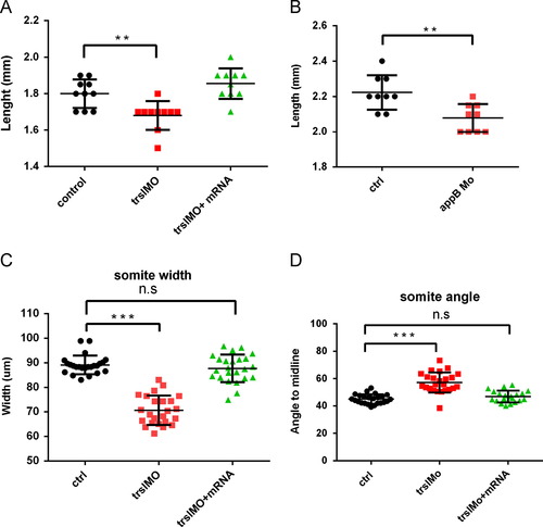
Changed body length and somite properties of embryos injected with trslMO or trslMO+mRNA. The average length of control (n=10), trslMO (n=10) and trslMO+mRNA (n=10) injected embryos at 24 hpf in embryo medium (A). Average length of control (n=10) and appbMO (n=10) embryos incubated with 0.02% MS-222 for 60 min (B). Measurement of the anterior to posterior somite width (C) and the somite-boundary to-midline-angle (C). Two-way ANOVA test: ***P<0.0005,**P<0.05. PHENOTYPE:
|

Locomotor behavior defects. Frequency of spontaneous coiling in appb morpholino (n=20, red) injected embryos and control (n=20, black) between 17 and 26 hpf (A). Total distance during spontaneous movement of embryos injected with control (black), trslMO (red) and trslMO+mRNA (green) between 22 and 72 hpf(B). Representative plot of movement (C). Startle duration of appbMO (n=15) and control MO (n=20) injected embryos at 3 dpf (D). PHENOTYPE:
|
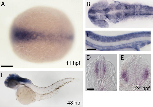
Expression of appb at 11, 24 and 48 hpf. The expression of appb is prominent in axial structures at 11 hpf (A) and is expressed in telencephalon, spinal cord neurons and pronephric duct and dorsal aorta by 24 hpf (B,C). Cross section at the caudal hindbrain and spinal cord shows expression in developing motor neurons and interneurons (D,E). The expression of appb becomes restricted to CNS at 48 hpf, (F). Scale bars: A, 100 μm; C, 80 μm; D, 40 μm. EXPRESSION / LABELING:
|
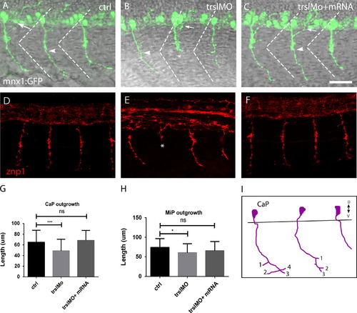
Defective primary motor neurons. PMNs in the spinal cord of control embryos (A), trslMO (B) and trslMO +mRNA (C) injected Tg(mnx1:GFP) embryos (B) have dorsally projecting MiPs (arrow) and ventrally projecting CaP (arrowhead). Staining of control (D), trslMO (E) and trslMO +mRNA (F) injected embryos using znp1. CaP neurons stalling was observed in trslMO injected embryos (N). Measurement of the length of CaP (G) and MiP (H). Bar is 20 μm. Two-way ANOVA test: NNNP<0.0005, NP<0.05, n.s.=not significant. |
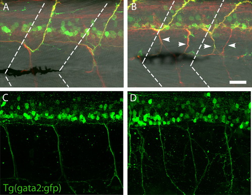
Impaired patterning of the secondary motor neurons. The Tg(isl1:GFP ) is expressed in SMNs of controls (A) but is reduced in appbMO injected embryos (B). Staining with the SMN-specific antibody zn5 shows irregular projections and stalled outgrowth of the SMN in appb morpholino injected embryos (B) compared to controls (A). The Tg(gata2:GFP) transgene shows that the irregular patterning of extending axons persist in morphants (D) while controls (C) have a regular pattern of SMN by 3 dpf. The dorsolateral fasciculus (DLF) which extends to the end of the spinal cord in controls (E) is projecting out to the tip of the tail fin in appb morphants (arrow, F). The contour of the tail fin is outlined by a dotted line. Bar: 20 μm. EXPRESSION / LABELING:
PHENOTYPE:
|
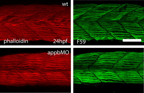
No change in the development of slow and fast muscle fibers. Staining of fast muscle fibers with phalloidin (A,C) and slow muscle fibers with F59 (B,D) does not reveal differences in muscle morphology or differentiation between control (n=20, A,B) and appbMO (n=23, C,D) injected fish. Scale bar is 20 μm. EXPRESSION / LABELING:
|
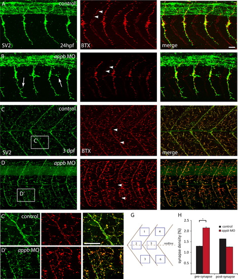
Increased pre-synapses and decreased post-synapses. Neuromuscular synapses are labeled with antibodies against SV2 (green, pre-synaptic vesicles) and αBTX (red, post-synaptic AChRs). Images are oriented so that left is rostral and top is dorsal. At 24 hpf, PMN of wildtype embryos (n=8) extends straight axons projecting dorsally and ventrally has established robust and overlapping NMJs (A). In appb morphants (n=8,) the ventral projections are branched (arrows) and have less post-synapses at the horizontal myoseptum (arrowheads) compared to control (B). At 3 dpf there is an elaborative network of pre- and post-synapses. Control larvae have well defined NMJs at the horizontal myoseptum (arrowheads) and evenly distributed NMJs in the somites (C). The appb morphants (D) have interrupted synapse formation at the horizontal myoseptum (arrowheads) and at the midline. (C′, D′) Higher magnification of C and D. (H) Measurement of the density of pre- and post-synapses were made by counting 6 areas (G) distributed dorsally, ventrally or at the midline of two somites of each fish. Scale bars: A, C 20 μm. EXPRESSION / LABELING:
PHENOTYPE:
|
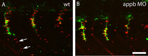
Lack of pre-formed AChR clusters. Clusters of AChR in the muscle were stained with fluorescently labeled αBTX (red, post-synaptic AChRs) and projecting motor neurons stained with SV2 (green, pre-synaptic vesicles) antibody. At 28 hpf, pre-formed clusters containing AChR (arrows) were found dorsal to the migrating motor neuron tip in controls (A) while these were absent in appbMO injected embryos (B). Scale bar: 20 μm. EXPRESSION / LABELING:
|
Reprinted from Developmental Biology, 381(2), Abramsson, A., Kettunen, P., Banote, R.K., Lott, E., Li, M., Arner, A., and Zetterberg, H., The zebrafish amyloid precursor protein-b is required for motor neuron guidance and synapse formation, 377-88, Copyright (2013) with permission from Elsevier. Full text @ Dev. Biol.