- Title
-
Iridophores and their interactions with other chromatophores are required for stripe formation in zebrafish
- Authors
- Frohnhöfer, H.G., Krauss, J., Maischein, H.M., and Nüsslein-Volhard, C.
- Source
- Full text @ Development
|
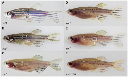
Adult phenotypes of iridophore mutants. (A) Wild-type fish. To denote individual stripes, we follow the nomenclature of Parichy et al. (Parichy et al., 2009), which we have extended by naming the interstripes, the central interstripe being X0. Additional stripes and interstripes added dorsally and ventrally during development are numbered according to their sequence of appearance. (B,C) rse mutants. Weak (B) and strong (C) rse alleles show a reduction of iridophores and melanophores. In strong rse mutants (Fig. 1C), the dense S-iridophore zone in X0 is lacking, and S-iridophores spread thinly from X0 dorsally and ventrally over the melanophores. In the weaker allele, a small ridge of dense S-iridopores persists (B). There are at most four melanophore stripes compared with five in a typical wild-type adult. Stripe 2D is reduced or absent, 2V is better preserved and 1V stays more strictly in a wild-type position parallel to X0 in rse compared with shd (D). (D,E) shd mutants. The white arrow in E displays an escaper S-iridophore patch (Lopes et al., 2008). In close proximity, the number of melanophores is significantly increased. (F) Double mutant rse;shd. The phenotype is indistinguishable from that of shd mutants. PHENOTYPE:
|
|
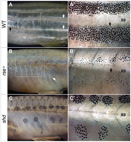
Details of adult phenotypes in iridophore mutants. Images of the anterior trunk of fixed animals photographed under white light. L-iridophores (black arrows) and dense S-iridophores (white arrows) reflect maximally at different angles of light and therefore shine up in different anteroposterior regions. Insets in A-C demarcate the magnified views in A′-C′ that were taken under UV light to avoid reflections from iridophores and to highlight xanthophores. Under this illumination, L-iridophores are visible as bundles of greyish vertically oriented fibres (black arrows). (A,A′) Wild-type fish. (B,B′) rse mutant fish. An illumination was chosen to avoid reflection of L-iridophores, whereas S-iridophores are visible (white arrow). L-iridophores, which are largely restricted to melanophore regions in wild type (A,A′, black arrows), expand into interstripe regions in rse mutants (B,B′). The light appearance in stripe 1D in B′ is due to the absence of L-iridophores. (C,C′) shd mutant fish. Ventrally, some of the melanophore patches (grey arrows) appear to be located between 1V and 2V. The light area in X0 in C′ is probably due to reflective properties of musculature. PHENOTYPE:
|
|
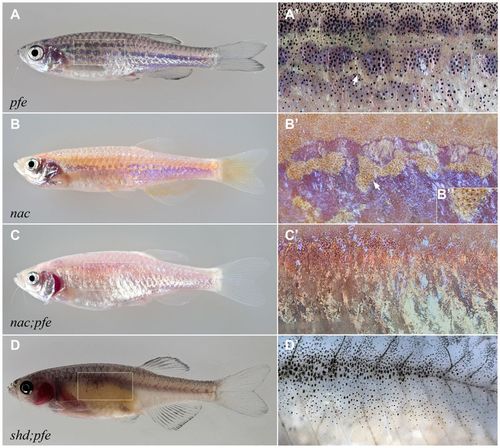
Adult phenotypes of xanthophore, melanophore and double mutants. pfe (A,A′), nac (B-B′), nac;pfe (C,C′) and shd;pfe (D,D′) mutant fish. The boxed areas in A-D are enlarged in A′-D′. Arrow in A′ marks X1V in the pfe mutant. In the nac mutant, xanthophores strictly colocalise with dense S-iridophores (B′); the arrow points to a region of X1V that is enlarged in B′. S-iridophores spread over the entire flank in nac;pfe (C′). Melanophores associated with the blood vessels are visible along myosepta in shd;pfe (D,D′). |
|
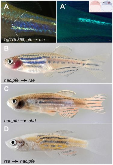
Phenotypes of chimeric animals. Pictures show stripes of wild-type pattern in chimeric animals produced by blastula cell transplantation of the indicated genotypes (A′, inset). The position of X0 is indicated. (A,A′) Tg(TDL358:gfp) → rse. Because GFP expression reduces with age, this animal displays weak labelling in X1V. The left boundary of the clone is marked with a white dashed line. (A) incident light, dark background, (A′) UV light. (B-D) The frequency of successful transplantations is highly variable, ranging from 10% (2/25, shd → nac;pfe; 7/41; rse → nac;pfe) to <50% (21/44 and 11/19 in transplantations of nac;pfe donors into rse and shd hosts, respectively). The size of the clones is also variable, but the quality does not depend on size, e.g. even in very small clones we observe the effect of iridophores on melanophore number. |
|
Phenotypes of chimeric animals. Pictures show stripes of wild-type pattern in chimeric animals produced by blastula cell transplantation of the indicated genotypes (A′, inset). The position of X0 is indicated. (A,A′) Tg(TDL358:gfp) → rse. Because GFP expression reduces with age, this animal displays weak labelling in X1V. The left boundary of the clone is marked with a white dashed line. (A) incident light, dark background, (A′) UV light. (B-D) The frequency of successful transplantations is highly variable, ranging from 10% (2/25, shd → nac;pfe; 7/41; rse → nac;pfe) to <50% (21/44 and 11/19 in transplantations of nac;pfe donors into rse and shd hosts, respectively). The size of the clones is also variable, but the quality does not depend on size, e.g. even in very small clones we observe the effect of iridophores on melanophore number. EXPRESSION / LABELING:
PHENOTYPE:
|
|
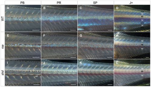
Iridophore development in the posterior trunk. (A-D) Wild type. Iridophores appear first at the horizontal myoseptum and grow in density. In the melanophore region, they maintain their bluish reflection, whereas their colour turns golden in the dense interstripe regions. Melanophores of the larval lateral stripe that are still present along the horizontal myoseptum (A) are cleared from this area during stages PR and SP (B,C). (E-H) rse mutant. Iridophores grow in number but remain always below the level of wild type. (I-L) shd mutant. No iridophores develop. Metamorphic melanophores avoid the region of X0, but the clearance of larval melanophores from this area is considerably delayed compared with wild type. ao aorta. Stages: PB, pelvic fin bud. PR, pelvic fin ray. SP, squamation onset posterior. J+, juvenile+. Scale bars: 250 μm. PHENOTYPE:
|
|
Iridophore development in the posterior trunk of xanthophore, melanophore and double mutants. pfe (A-D), nac (E-H), nac;pfe (I-L) and shd;pfe (M-P) mutants. In pfe, as well as in nac and nac;pfe mutants, X0 is more prominent than in wild type; it expands to cover the entire flank in nac;pfe mutants. In shd;pfe mutants, melanophores distribute evenly also in the territory normally occupied by X0. Stages: PB, pelvic fin bud; PR, pelvic fin ray. SP, squamation onset posterior; J+, juvenile+. Scale bars: 250 μm. PHENOTYPE:
|
|
The choker phenotype. (A,B) Adult individuals, displaying parallel stripes with arbitrary orientation. choker adults can reach normal sizes and are fertile. (C-E) Metamorphic development. In the absence of the horizontal myoseptum, melanophores appear before iridophores and xanthophores, evenly dispersed over the flank (C). Iridophores and xanthophores emerge in patches interspersed with melanophores (D), but then aggregate into stripe-like arrangements, and separate from melanophore regions (E). The aorta is visible as dark horizontal shade in C. Scale bars: 250 μm. PHENOTYPE:
|
|
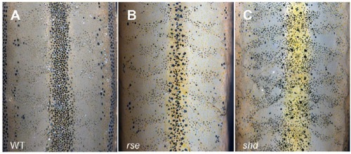
Dorsal view of wild type and iridophore mutants. Wild-type (A), rse (B) and shd (C) adult fish. In the mutants, the number of melanophores in the stripe along the dorsal midline is reduced, and they fail to aggregate properly. Anterior is top. Scales are removed, but many small epidermal melanophores of the scale pockets are still visible. The larger melanophores directly at the midline are dermal. |

