
- Title
-
Characterization and expression analysis of mcoln1.1 and mcoln1.2, the putative zebrafish co-orthologs of the gene responsible for human mucolipidosis type IV
- Authors
- Benini, A., Bozzato, A., Mantovanelli, S., Calvarini, L., Giacopuzzi, E., Bresciani, R., Moleri, S., Zizioli, D., Beltrame, M., and Borsani, G.
- Source
- Full text @ Int. J. Dev. Biol.

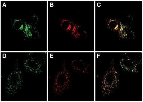
Subcellular localization of zebrafish Mcoln1.1 (A) and Mcoln1.2 (D) proteins transiently expressed in HeLa cells. LAMP1 positive vesicles are shown in (B) and (E). A partial co-localization of both zebrafish proteins was observed in LAMP1-positive vesicles (C) and (F). |
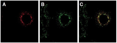
Subcellular localization of zebrafish Mcoln1.1 (A) and Mcoln1.2 (D) polypeptides as well as human TRPML1 (B,E) in transiently transfected HeLa cells. Co-localizations between the human and the zebrafish polypeptides are shown in (C) and (F). |
Transient coexpression of Mcoln1.1 (A) and Mcoln1.2 (B) in HeLa cells reveals a partial co-localization of the two polypeptides (C). |

Real Time PCR expression analysis of mcoln1.1 and mcoln1.2 genes throughout Danio rerio development. All reactions were run in triplicate. The relative expression levels, represented as the mean(SEM in log2 scale, were determined with respect to the 1-cell stage and normalized to elongation factor 1α (ef1α). |

RT-PCR expression analysis of mcoln1.1 and mcoln1.2 in adult zebrafish tissues. Beta-actin was also amplified as housekeeping gene internal control. 1: brain; 2: intestine; 3: eye; 4: heart; 5: kidney; 6: swim bladder; 7: branchias; 8: testis; 9: ovary; 10: negative control. A densitometric analysis of RT-PCR bands is reported below each panel. The units at the left of the graphs are arbitrary. EXPRESSION / LABELING:
|
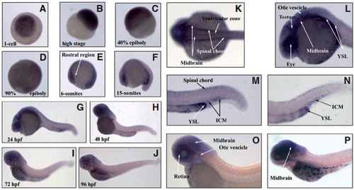
(A-J) Whole-mount in situ hybridization of mcoln1.1 at different stages of zebrafish development (63X magnification). Dorsal (K) and lateral (L) view of 24 hpf embryo head. Magnification of 24 hpf (M) and 36 hpf (N) embryo tails. Lateral view of 48 hpf (O) and 80 hpf (P) embryo. Images (K-P) were acquired at 115X magnification. Relevant sites of mcoln1.1 gene expression are indicated with arrows. |

(A-J) Whole-mount in situ hybridization of mcoln1.2 transcripts during zebrafish development (63X magnification). Dorsal (K) and lateral (L, head; M, tail) view of 24 hpf embryo. Lateral (N, tail; P, head) and dorsal (O) view of 36 hpf embryo. Dorsal view at 72 hpf (Q). High magnification of the eye at 96 hpf (R). Images (K-R) were acquired at 115X magnification. Relevant sites of mcoln1.2 gene expression are indicated with arrows. |