- Title
-
Zebrafish model for allogeneic hematopoietic cell transplantation not requiring preconditioning
- Authors
- Hess, I., Iwanami, N., Schorpp, M., and Boehm, T.
- Source
- Full text @ Proc. Natl. Acad. Sci. USA
|
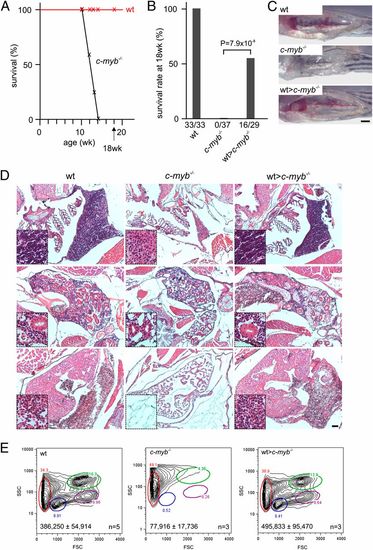
Reconstitution of the c-myb-/- mutant phenotype. (A) Survival of wild-type (n = 33) and c-myb-/- mutant (n = 37) fish; mutants do not survive longer than 14 wk of age. (B) Survival rates of wild-type, c-myb-/- mutants and mutant recipients transplanted with wild-type WKM (wt>c-myb-/-) transgenic for an ikaros:eGFP reporter assessed at 18 wk of age. All deaths occurring within the first 24 h after transplantation were considered to be procedural and thus not used for the calculation of survival rates. (C) Macroscopic appearance of head kidneys in wild-type, c-myb/ mutants and transplanted mutant recipients; note that mutant kidney shows a complete lack of hematopoietic cells (17). (Scale bar, 1 mm.) (D) Hematoxylin/eosin staining of sections through the region of the thymus (Top), kidney (Middle), and heart (Bottom) of wild-type, c-myb-/- mutants and transplanted recipients at 9 wk of age (i.e., 3 wk after transplantation); insets are at fourfold higher magnifications. Note the lack of hematopoietic cells in the mutants and complete hematopoietic reconstitution in transplanted recipients. (Scale bar, 50 μm.) (E) Flow cytometric analyses of WKM cells of wild-type, c-myb-/- mutants and transplanted recipients as in D. FSC, forward light scatter; SSC, side light scatter. Circles denote different blood cell populations in adult wild-type fish: red, erythrocytes; blue, lymphocytes; magenta, precursors; green, myelomonocytes. Total cell numbers (mean± SD) in WKM preparations are indicated. PHENOTYPE:
|
|
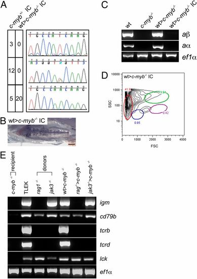
Characterization of reconstituted c-myb-/- mutants. (A) Genotypes of offspring embryos resulting from in-crosses of c-myb+/- heterozygous fish and reconstituted female and male c-myb-/- mutant fish, respectively. The fecundity of reconstituted transplant recipients is indistinguishable from wild-type fish. The number of wild-type (Top panels), heterozygous (Middle panels), and homozygous mutant (Bottom panels) embryos are indicated as are representative sequence traces. (B) Macroscopic phenotype of the head kidney in the offspring of reconstituted c-myb-/- mutants. (Scale bar, 1 mm.) (C) Expression of adult hemoglobin alpha (aα) and beta (aβ) genes in WKM cells of wild-type (wt), c-myb-/- mutants; reconstituted mutants (wt>c-myb-/-); and offspring of reconstituted mutants (wt>c-myb-/- IC) as determined by RT-PCR at 11–12 wk of age; amplification with ef1α-specific primers serves as a control for cDNA integrity and amount. (D) Flow cytometry analysis of WKM cells from offspring of reconstituted mutant fish (see Fig. 1E legend for explanation of indicated cell populations); profile representative of three fish. (E) Expression of T- and B-cell–specific marker genes in TLEK fish (wild-type strain), c-myb-/- mutants, rag1- and jak3-deficient donor fish, and c-myb-/- recipients transplanted with wild-type (transgenic for the ikaros:eGFP reporter), rag1-, and jak3-deficient WKM cells. cd79b, gene encoding the igβ adaptor protein, constituting part of the B-cell receptor in B cells; igm, Ig μ heavy chain gene expressed in B cells; lck, gene encoding a receptor-associated tyrosine kinase expressed in T cells; tcrb, T-cell receptor β chain gene expressed in T cells; tcrd, T-cell receptor δ chain gene expressed in T cells. |
|
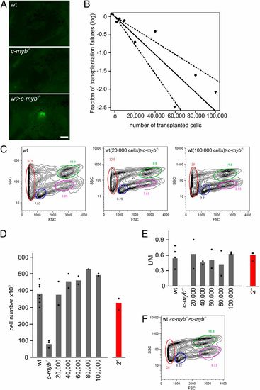
Long-term multilineage reconstitution of c-myb-/- mutant fish. (A) Thymus colonization in c-myb-/- recipients transplanted with wild-type ikaros:eGFP-transgenic WKM cells. Photographs (side views) were taken 7 d after transplantation. (Scale bar, 50 μm.) (B) Limiting dilution analysis of hematopoietic reconstitution of c-myb-/- mutant fish. (C) Flow cytometric analysis of WKM of wild-type fish (Left) and stably reconstituted c-myb-/- mutants after transplantation of 20,000 (Middle) and 100,000 (Right) wild-type WKM cells (see Fig. 1E legend for explanation of indicated cell populations). (D) Cellularity of WKM of wild-type (wt) fish, c-myb-/- mutants, and mutants stably reconstituted after transfer of the indicated numbers of WKM wild-type cells (9 wk after transplantation). The mean values (bars) and results of individual fish are shown. The number of cells found in WKM in secondary transplant recipients (2°) is depicted in red. (E) Multilineage reconstitution as measured by the ratio of cells in lymphoid (L) and myeloid (M) gates in flow cytometric analyses for fish depicted in D. (F) Flow cytometric analysis of WKM of stably reconstituted c-myb-/- mutants after transplantation of WKM of primary recipients (see Fig. 1E legend for explanation of indicated cell populations). |
|
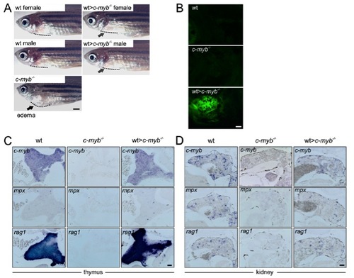
Characterization of reconstituted c-myb-/- mutants. (A) Side views of wild-type, c-myb-/- transplant recipients and a c-myb-/- mutant fish. Note the absence of cardiac edema (resulting in a bulged body curvature) in the transplanted fish; the body curvature is highlighted by dotted lines. (Scale bar, 1 mm.) (B) Reconstitution of T-cell development in the thymus of c-myb-/- mutant recipients transplanted with ikaros:eGFP-transgenic wild-type whole kidney marrow (WKM) cells; note the presence of green fluorescent cells in the transplanted fish. Photographs (side views) were taken 20 d after transplantation. (Scale bar, 50 μm.) (C and D) Whole-mount RNA in situ hybridization with c-myb-, mpx-, and rag-specific probes on thymus (C) and kidney (D) sections of wild-type, c-myb-/- mutant and c-myb-/- transplant recipients (3 wk after transplantation). In mutant tissues, no positive cells were observed for c-myb, mpx, and rag1, while expression patterns in transplanted c-myb-/- recipients are indistinguishable from those of their wild-type siblings. c-myb is a general marker for hematopoietic cells; mpx is a marker for myeloid cells; rag1 is a marker for developing lymphocytes. (Scale bar, 50 μm.) PHENOTYPE:
|


