
- Title
-
Zebrafish Mnx proteins specify one motoneuron subtype and suppress acquisition of interneuron characteristics
- Authors
- Seredick, S., Van Ryswyk, L., Hutchinson, S.A., and Eisen, J.S.
- Source
- Full text @ Neural Dev.

Zebrafish mnx family genes are dynamically expressed in PMNs and VeLD interneurons. Lateral views, anterior to the left; all figures are in this orientation unless otherwise noted. MiPs and RoPs are outlined green in panels with islet1 labeling, CaPs and VaPs are outlined blue in panels with islet2a labeling. Segment boundaries are demarcated with diagonal lines. (A-L) RNA expression; two spinal hemisegments are shown in each panel. At 14 hpf, mnx1 is co-expressed with both islet1 (A) and islet2a (B), mnx2a is only co-expressed with islet2a (E, F), and mnx2b is co-expressed with both islet1 (I) and islet2a (J). The VeLD interneuron can be seen expressing both mnx1 (arrowheads A, B) and mnx2b (arrowheads I, J). At 24 hpf, mnx1 maintains co-expression with both islet1 (C) and islet2a (D), mnx2a is co-expressed with both islet1 (G) and islet2a (H), and mnx2b is co-expressed only with islet1 (K, L). VeLD still expresses mnx1 (arrowhead C). (M-N) Protein expression. Mnx2b protein (red) co-localizes with the MiP soma (GFP; green) (M). Mnx1 protein (red) co-localizes with the VeLD soma (GFP; green) (N). (O) Schematic of mnx expression. Scale bar: 10 μm, A-L; 10 μmm M; 20 μm N. EXPRESSION / LABELING:
|

Mnx proteins are not required for formation of PMNs or VeLDs. Control and mnx MO-injected embryos labeled with markers of PMN or VeLD identity. (A-B) At 16 hpf, PMNs express chat and nrp1a:GFP in control (A) and mnx MO-injected embryos (B). (C-D) At 18 hpf, PMNs express Islet in control (C) and mnx MO-injected Tg(nrp1a:GFP) embryos (D). (E-F) At 20 hpf, VeLD interneuron (arrowheads) numbers and distribution are the same in control (E) and mnx MO-injected Tg(vsx1:GFP) embryos (F); n = 80 segments in 10 embryos for control and mnx MO-injected samples. Scale bar: 15 μm, A-B; 20 μm, C-F. EXPRESSION / LABELING:
|

Mnx proteins promote MiP subtype identity and prevent interneuron-like and CaP-like processes. (A-D) Control and mnx MO-injected embryos. (A) In controls islet1 and islet2a expression is mutually exclusive at 18 hpf. (B) In embryos injected with mnx1, mnx2a, and mnx2b MOs, islet1 is co-expressed with islet2a in CaP and VaP. (C) Control embryos have normal, ventrally-projecting CaP axons (arrows) and dorsally-projecting MiP axons (arrowheads). (D) CaP axons (arrows) are normal in mnx MO-injected embryos whereas MiP axons (closed arrowheads) are absent and there are ectopic interneuron-like axons (open arrowheads). (E-J′′) Rhodamine-dextran-labeled MiPs in 28 to 32 hpf Tg(nrp1a:GFP) embryos injected with mnx1, mnx2a, and mnx2b MOs. Panels show rhodamine-dextran labeling (no superscript), nrp1a:GFP (2) and a merged image of the two channels (′′). Labeled MiPs (yellow arrowheads) become hybrids with six morphologies. (E-E′′) V2a hybrids express nrp1a:GFP and have a descending V2a-like axon. (F-F′′) MiP-V2a hybrids have a descending V2a-like axon as well as a normal-appearing MiP ventral axon; the red blob to the right is a cell that was killed during labeling. (G-G′′) MiP-V2a-CaP hybrids have a descending V2a-like axon as well as a ventrally-projecting CaP-like axon. (H-H′′) MiP-CaP hybrids have both a normal MiP axon and a CaP-like axon. (I-I′′) Truncated MiP-CaP hybrids have a truncated MiP dorsal axon as well as a ventrally-projecting CaP-like axon. (J-J′′) CaP hybrids have a CaP-like axon that extends next to a normal CaP axon. Scale bar: 20 μm, A-D; 40 μm, E-H′′. EXPRESSION / LABELING:
PHENOTYPE:
|
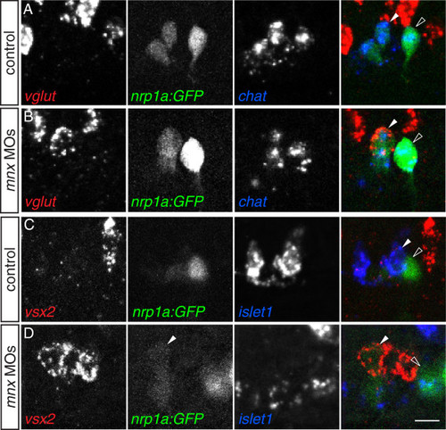
Mnx proteins prevent MiPs from acquiring V2a-like molecular characteristics. (A-D) Single spinal hemisegments of control and mnx MO-injected embryos. MiPs (closed arrowheads) and CaPs (open arrowheads) are indicated in merged panels (right column). (A) In control embryos, MiPs and CaPs express chat but never vglut (0/20 vglut+ MiPs or CaPs in five embryos). (B) In mnx MO-injected embryos, MiPs co-express both chat and vglut (22/41 vglut+ MiPs in eight embryos), while CaPs express chat but not vglut (0/41 vglut+ CaPs in eight embryos. (C) In control embryos, MiPs express islet1 but not vsx2 (0/50 vsx2+ MiPs in 10 embryos), while CaPs express neither islet1 nor vsx2 (0/50 vsx2+ CaPs in 10 embryos). (D) In mnx MO-injected embryos, MiP express vsx2 but not islet1 (26/60 vsx2+ MiPs in 13 embryos) while CaPs express neither vsx2 nor islet1 (0/60 vsx2+ CaPs in 13 embryos). Scale bar: 10 μm. EXPRESSION / LABELING:
|
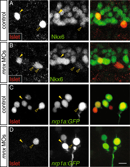
Mnx proteins maintain the late phase of Islet1 expression in MiPs independently of Nkx6. (A-D) Single spinal hemisegments of control and mnx MO-injected embryos. MiPs (closed arrowheads) and CaPs (open arrowheads) are indicated in each of the single channel panels; right column shows merged channels. (A) At 17 hpf, MiPs in control embryos strongly express Islet and Nkx6 (17/30 Nkx6+ MiPs in seven embryos). CaPs strongly express Islet but weakly express Nkx6. (B) MiPs in mnx MO-injected embryos continue to strongly express Islet and Nkx6 (13/22 Nkx6+ MiPs in five embryos). (C) At 21 hpf in Tg(nrp1a:GFP) control embryos, both MiPs and CaPs express Islet (32/32 Islet+ CaPs, and 31/32 Islet+ in five embryos. (D) In mnx MO-injected Tg(nrp1a:GFP) embryos, CaPs (60/60 Islet+ CaPs in 10 embryos) but not MiPs (19/60 Islet+ MiPs in 10 embryos), strongly express Islet. Scale bar: 20 μm. EXPRESSION / LABELING:
|

mnx1 and mnx2b are both expressed in VeLD interneurons. (A-D) VeLD somata are outlined. (A) At 16 hpf, mnx1 and mnx2b are co-expressed in VeLD. (B) At 24 hpf, mnx1+ VeLDs express gad. (C, D) At 24 hpf mnx1+ VeLDs express neither vsx2 (C) nor vglut (D). Scale bar: 10 μm. |

Mnx proteins are restricted to post-mitotic neurons. Lateral views of 12 to 14 hpf Tg(olig2:GFP) embryos. (A) Mnx2a+ cells within the spinal cord do not co-express phosphohistone H3, a marker of mitotic cells (0/153 Mnx2a+ cells in 13 embryos). Some Mnx2a+ cells strongly express GFP. (B) Mnx1+ cells within the spinal cord do not co-express phosphohistone H3 (0/70 Mnx1+ cells in 10 embryos). No Mnx1+ cells strongly express GFP. (C) Mnx2a+ cells that expressed GFP weakly or were GFP- co-expressed Elavl3, a marker of post-mitotic neurons. Mnx2a+ cells that expressed GFP strongly did not co-express Elavl3. (D) Mnx1+ cells co-expressed Elavl3. (E) Schematic of gene expression during transition from pMN progenitors to post-mitotic neurons. Mitotic progenitors express phosphohistone H3 (PH3), whereas post-mitotic neurons express Elavl3. olig2 expression is initiated in progenitors, and down-regulated as cells become post-mitotic. Both Mnx1 and Mnx2a expression is initiated after cells become postmitotic, with expression of Mnx2a preceding expression of Mnx1. Scale bar: 30 μm, A-D. |

Mnx proteins are differentially expressed in secondary motoneurons. (A-D) Protein expression in two spinal hemisegments of 26 hpf Tg(mnx1:GFP) embryos. The single channel panels show Mnx protein expression. (A, B) Mnx1 is strongly expressed by PMNs and VeLD interneurons, and more weakly expressed by a subset of mostly dorsally-located secondary motoneurons. (C, D) Mnx2a appears to be down-regulated in PMNs and is expressed by a subset of mostly ventrally-located SMNs. Scale bar: 20 μm. |

With the exception of mnx2b which is regulated by Lhx3 and Lhx4, expression of mnx genes is independent of Islet, Nkx6, Lhx3, Lhx4, and other Mnx paralogs. (AAA) Lateral views of control and MO-injected embryos colabeled with islet2a (green) to mark CaP and VaP. Segment boundaries are demarcated with diagonal lines. At 18 hpf, expression of mnx1 (A, B), mnx2a (C, D) and mnx2b (E, F) are unaffected by absence of Islet1. Note that islet2a is not expressed in the absence of islet1. At 18 hpf, expression of mnx1 (A, G), mnx2a (C, H) and mnx2b (E, I) are unaffected by absence of Nkx6.1 and Nkx6.2. At 24 hpf, expression of mnx1 (J, K) and mnx2a (L, M) are unaffected by absence of Lhx3 and Lhx4. mnx2b is not expressed in the absence of Lhx3 and Lhx4 (N, O). At 18 hpf, expression of lhx3 (P, Q) and lhx4 (R, S) are unaffected by absence of Mnx proteins. At 16 hpf, expression of mnx2a (T, U) and mnx2b (V, W) are unaffected by absence of Mnx1. (XAA) Embryos labeled only for expression of mnx genes. Expression of mnx1 (X, Y) and mnx2b (Z, AA) are unaffected by absence of Mnx2a. We did not examine expression of mnx1 and mnx2a in the absence of Mnx2b as we did not detect Mnx2b protein before 20 hpf. Note that panels M, O and Q are reproduced from Figure 1 to facilitate comparison of gene expression in control and MO-injected embryos. Scale bar: 10 μm. |

Morpholinos targeting mnx family genes are specific and effective in knocking down protein. Lateral views of two spinal hemisegments, segment boundaries denoted by diagonal lines, of uninjected and control MO-injected Tg(mnx1:GFP) embryos labeled for antibodies against Mnx1 (A, D), Mnx2a (B, E), and Mnx2b (C, F). Embryos injected with mnx1 MO lack Mnx1 antibody labeling (G), but maintain Mnx2a (H) and Mnx2b (I) antibody labeling. Embryos injected with mnx2a MO lack Mnx2a antibody labeling (K), but maintain Mnx1 (J) and Mnx2b (L) antibody labeling. Embryos injected with mnx2b MO lack Mnx2b antibody labeling (O), but maintain Mnx1 (M) and Mnx2a (N) antibody labeling. Scale bar: 20 μm. |

In the absence of Mnx proteins, neither MiPs nor CaPs aberrantly express GABAergic or glycinergic markers. (A-D) Lateral views of single hemisegments of control and mnx MO-injected embryos. MiPs (closed arrowheads) and CaPs (open arrowheads) are indicated in merged panels (column on right). (A, B) In control and MO-injected embryos, MiP and CaP express chat but not gads. (C, D) In control and MO-injected embryos, MiP and CaP express chat but not glyts. Scale bar: 20 μm. |