
- Title
-
Structural disorganization of pronephric glomerulus in zebrafish mpp5a/nagie oko mutant
- Authors
- Ichimura, K., Fukuyo, Y., Nakamura, T., Powell, R., Sakai, T., and Obara, T.
- Source
- Full text @ Dev. Dyn.

Mpp5a protein localization in zebrafish pronephric glomerulus. Immunoreactivity for Mpp5a protein is found in both medial and lateral half of nephron primordia at 34 hpf (arrowhead and small arrows in A). At 3 dpf, the signal for Mpp5a is found along the glomerular capillary wall as a tortuous line (arrowhead in B), in addition to the pronephric tubule (small arrows in B). Mpp5a signal is also found at the position of apical tight junction of neural tube (large arrows in A, B). In 3 dpf, immunoreactivity for ZO-1 (red) and Mpp5a (green) is colocalized within the glomerulus (arrowheads in C–C′′). Mpp5a signal is also found at parietal epithelium of Bowman′s capsule (arrows in C–C′′). In, intestine; NC, notochord. Scale bar = 10 μm. EXPRESSION / LABELING:
|

mpp5am520 mutant displays structural disorganization of pronephric glomerulus. Pronephric glomerular structure is shown by hematoxylin-eosin stained JB-4 section at 2 dpf (A-C), 3 dpf (D–F), and 4 dpf (G-I). In wild type siblings, a pair of glomerular primordia has merged to form a single glomerulus (arrowheads) beneath the notochord at 2 dpf (A), 3 dpf (D), and 4 dpf (G). In 2-dpf mpp5am520 mutants, a pair of glomerular primordia retains epithelial vesicular structure that does not merge beneath the notochord (arrows in B, C). At 3 and 4 dpf mpp5am520 mutants still exhibited two separated podocyte masses (arrows in E, F, H, I). Scale bar = 10 μm. |
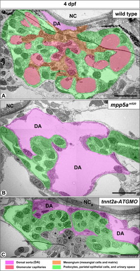
mpp5am520 mutants and tnnt2a-ATG morphants display a similar structural disorganization of pronephric glomerulus. A: In 4-dpf wild-type embryos, the pronephric glomerulus contains several glomerular capillaries together with mesangium, which is arborized in shape. Podocytes and GBM cover the glomerular capillaries and mesangium en bloc. In both 4-dpf mpp5am520 mutants (B) and tnnt2a-ATG morphants (C), the extremely dilated dorsal aorta (DA) lies between a pair of glomerular primordia, which do not contain any fine glomerular capillaries or mesangium. NC, notochord. Scale bar = 500 nm. PHENOTYPE:
|
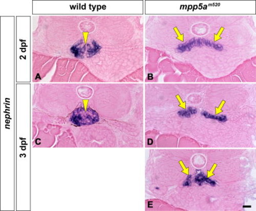
nephrin mRNA expression in pronephric glomeruli of mpp5am520 mutant. Position and shape of pronephric glomerulus are shown by in situ hybridization for nephrin mRNA at 2 dpf (A, B) and 3 dpf (C-E), which only express in podocytes. nephrin mRNA is expressed in the glomerulus at 2 and 3 dpf in wild-type embryos as a single globular mass beneath the notochord (A, C; arrowheads). In the mpp5am520 mutant, the glomerulus (glomerular primordia) express nephrin mRNA in a domain shaped like a moustache at 2 and 3 dpf (arrows in B, D, E). Scale bar = 10 μm. EXPRESSION / LABELING:
PHENOTYPE:
|
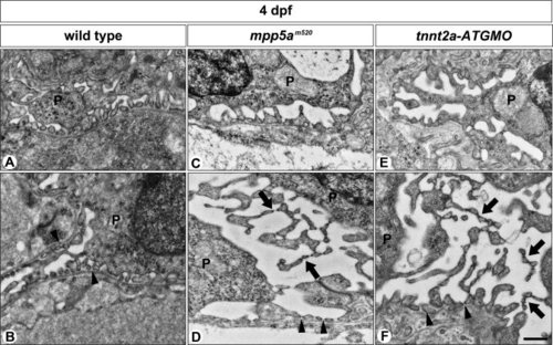
Podocyte foot processes are formed in mpp5am520 mutants and tnnt2a-ATG morphants. A, B: In wild-type siblings, foot processes with SD are vigorously formed by 4 dpf. In 4-dpf mpp5am520 mutants (C, D) and tnnt2a-ATG morphants (E, F) podocytes form regular foot processes with SD by 4 dpf as observed in the wild-type siblings. In most podocytes, multiple microvillus-like processes have protruded from the apical surface of the cell body and extend into the Bowman′s space (arrows in D, F). In addition, they form regular foot processes adhering to the GBM (arrowheads in B, D, F). These microvillus-like processes are connected to each other by SD-like structures. P, podocyte. Scale bar = 500 nm. PHENOTYPE:
|

A tnnt2a-ATG morphant displays glomerulus disorganization similar to that found in the mpp5am520 mutants. Pronephric glomerular structure and nephrin mRNA expression are shown by hematoxylin-eosin stained section (A-F) and in situ hybridization with eosin-stained (G-J), respectively. In wild-type (A, C, E, G, I), a pair of glomerular primordia has already merged to form a single glomerulus (arrowheads) beneath the notochord at 2 dpf (A, G), 3 dpf (C, I), and 4 dpf (E). In tnnt2a-ATG morphants (B, D, F, H, J), a pair of glomerular primordia has retained their epithelial vesicular structure and remains unmerged at the midline in 2 dpf (arrows in B, H). Glomerular primordia have still not merged, and an extremely dilated dorsal aorta is interposed between glomerular primordia at 3 dpf (D) and 4 dpf (F). Bowman′s space is apparent in most cases of tnnt2a-ATG morphants (D, F). nephrin-expressing regions are moustache-like in shape and expanded in the direction of the pronephric tubule (arrows in H, I), as is the case in the mpp5a mutants. Scale bar = 10 μm. EXPRESSION / LABELING:
PHENOTYPE:
|

Unillustrated author statements PHENOTYPE:
|