
- Title
-
Multilayer mounting enables long-term imaging of zebrafish development in a light sheet microscope
- Authors
- Kaufmann, A., Mickoleit, M., Weber, M., and Huisken, J.
- Source
- Full text @ Development

Influence of different mounting media on growth and immobility. (A,A′) Zebrafish embryo [Tg(myl7:GFP)] embedded in 1.5% agarose from 24-48 hpf exhibits a short crooked tail (arrowhead) and disturbed morphology of the heart (arrow). (B) Immobility, heart rate and edema after incubation from 24-72 hpf in different concentrations of Tricaine. Superimposed time-lapse images of one embryo are shown for four concentrations (pictograms 1-4). 0% immobility refers to movement in E3 (control); 100% immobility corresponds to 200 mg/l Tricaine. Tricaine concentrations as low as 133 mg/l result in immobilization of the fish without altering the heart rate. Edemas were observed with concentrations as low as 100 mg/l. (C) Immobility and growth in different agarose concentrations after embedding embryos from 24-72 hpf. At 0.1% agarose, both immobility and normal growth were achieved. (C′) Immobility and growth in different methylcellulose concentrations after embedding embryos from 24-72 hpf. At 2.5-3.0% methylcellulose, both immobility and growth were satisfactory. Mean ± s.d., n=6. Dashed lines represent empirical fits for better visualization. |

Influence of tube enclosure on growth. (A) Kinetics of embryonic growth during 2 days in E3, 0.1% agarose, 3.0% methylcellulose, with and without FEP tubes. (B,C) Length (B) and morphology (C) of 72-hpf zebrafish embryos from A. Mean ± s.d., n=6. *, P<0.01. |

Optical quality of FEP mounting. (A) Schematic of mounting in 1.5% agarose (left) and multilayer mounting in 0.1% agarose in coated FEP tubes (right). (B) Principle of SPIM and direction of axes for the analysis of optical quality. Not to scale. (C) Maximum intensity projections of averaged images of beads (n, number of beads) in 1.5% agarose without enclosure, in an FEP tube and in a glass capillary. (D) Full width at half maximum (FWHM). Mean ± s.d., n=6. (E) Developing zebrafish embryo [Tg(kdrl:GFP)] embedded in 0.1% agarose in a coated FEP tube during SPIM time-lapse (supplementary material Movie 1). Images resolve even the finest structures, e.g. the cell extensions forming the parachordal vessels (arrowheads). |
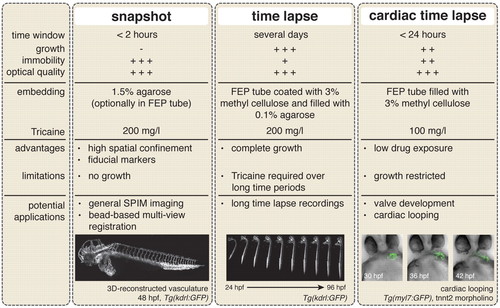
Recommended mounting protocols for light sheet microscopy. Three different experimental settings and their potential applications. The image of the 3D-reconstructed vasculature is a maximum projection (supplementary material Movie 2). Images of the developing zebrafish embryo in 0.1% agarose and the developing heart in 3.0% methylcellulose are taken from supplementary material Movies 1 and 3, respectively. |
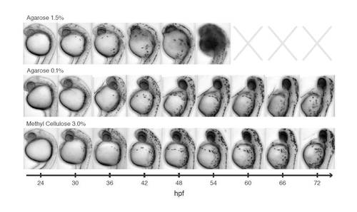
Influence of mounting conditions on morphological development. Embryos were embedded in 1.5% agarose, 0.1% agarose in an FEP tube, or 3.0% methylcellulose in an FEP tube at 24 hpf and imaged in transmission with SPIM. Images were taken every 6 hours. The embryo embedded in 1.5% agarose is unable to undergo morphological changes and died at 50 hpf. Multilayer mounting methods allow outgrowth of the head. |