
- Title
-
Developmental Role of Zebrafish Protease-Activated Receptor 1 (PAR1) in the Cardio-Vascular System
- Authors
- Ellertsdottir, E., Berthold, P.R, Bouzaffour, M., Dufourcq, P., Trayer, V., Gauron, C., Vriz, S., Affolter, M., and Rampon, C.
- Source
- Full text @ PLoS One
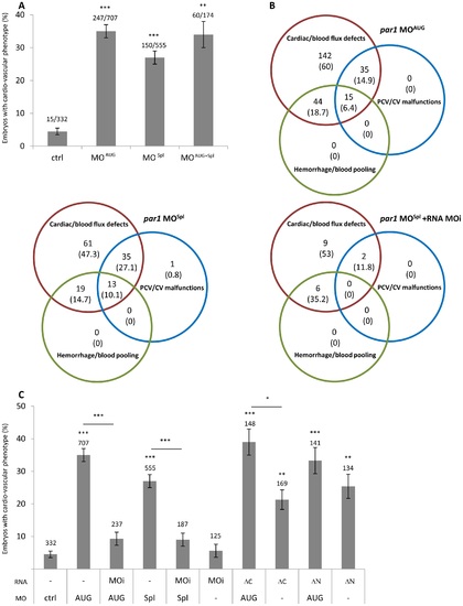
par1 knockdown induces cardio-vascular phenotype at 48 hpf. (A) cardio-vascular phenotype in control and par1 morphants. n values were indicated on the top of each bar of the graph. (B) Venn diagram representation of three classes of vascular failure. n values (% of embryos/embryos with cardio-vascular phenotype) were indicated. (C) cardio-vascular phenotype of morpholino and/or RNA injected embryos. Three par1 mRNA mutants were used. n values were added on the top of each bar of the graph. Average ± error bars from ≥3 independent experiments are presented. * p<0.05; ** p<0.01; *** p<0.001. ctrl, control; MOAUG, par1 MOAUG; MOSpl, par1 MOSpl; MOi, par1 mRNA morpholino-insensitive; ΔC, par1 mRNA morpholino-insensitive lacking intracellular domain; ΔN, par1 mRNA N terminus deleted. PHENOTYPE:
|

Slow heart rate in par1 morphants. (A) Heart rates were determined at 48 hpf and significance was described from an ANOVA-test statistic. The heart rate of par1 morphants was significantly lower than controls. *** p<0.001. n values were indicated. (B) Classification of heart rate. Class 1: heart shows no heart rate; class 2: heart presents lower heart rate <90 bpm; class 3: heart rate seems normal >90 bpm. (C) Ventricular shortening fraction (D) The heart is labeled with anti-myosin heavy chain antibody in 48 hpf embryos. Ventral view with the head on the top. a, atrium; v, ventricle, L; embryo left. PHENOTYPE:
|
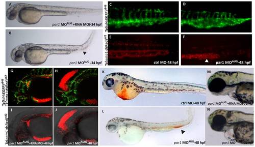
par1 knockdown causes ICM blood pooling, heart edema and head hemorrhage. (A) 34 hpf rescued embryo with heart beat and blood flow, resembling a ctrl MO embryo at this stage. (B) par1 morphant showing blood pooling (arrowhead) with bend tail and heart edema. (C–F) The posterior part of the embryo at 48 hpf. The ctrl MO embryo has dorsal aorta, CV, ISVs and arteries with expanded lumen with blood flow as seen in the double transgenic background (C and E). The par1 morphant shows ISVs, arteries, dorsal aorta and CV, but the blood cells are pooled in the CV (arrowhead in F) and the vein is bulged and malformed (asterisk in D). (G–J) Head of 48 hpf double transgenic embryos, displaying endothelial cells in green fluorescence and blood cells in red fluorescence (Tg(kdrl:EGFP)s843; Tg(gata1:dsRed)sd2). The blood vessels in the head of both the rescued embryo and the morphant are developed normally, but there is a leakage of blood cells into the mesencephalon of the morphant embryo, causing hemorrhage. Blood flow in the body of both rescued and morphant embryo was normal (not shown). (K–L) O-dianisidine staining at 48 hpf. (K) embryo treated with Ctrl MO displaying normal blood circulation; (L) embryo treated with par1-MOAUG showing reduced level of Hb in the Duct of Cuvier and blood pooling in the tail. (M–N) Embryos at protruding mouth stage (72 hpf). Contrary to the rescue embryo (M), the par1 morphant has a slight cardiac edema, no mouth protrusion and no visible swim bladder (N). PHENOTYPE:
|
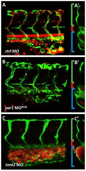
par1 knockdown impairs vascular remodeling in the cardinal vein. (A–B) Lateral views of double transgenic embryos (Tg(kdrl:EGFP)s843;Tg(gata1:dsRed)sd2) at 35 hpf. (A) The intersegmental vessels are connected to the CV and the one intersegmental artery (most posterior) seen in the figure has blood flowing from the dorsal aorta. (A2) In the mid-cross section, the intersegmental artery is visible (green bracket), the dorsal aorta can be seen as one tube (red bracket), and the CV is seen as two tubes (blue bracket). (B) par1 morphant lateral view. ISVs are lumenized (asterisk) (B2) In cross section, the CV tube does not show a defined number. Due to mislaid vein remodeling the tube is disorganized, cell clusters are apparent instead of a round tube. (C) The tnnt2 knockdown shows no mature inflation of the dorsal aorta and only a minor primary lumen in the ISV. The CV is bulged and full of blood cells in this region. (C′) The small dorsal aorta is a clear difference to the par1 knockdown, additionally to the lack of lumen in the ISVs and blood cells always clogging in the region of origin, PBI. PHENOTYPE:
|

The endothelial adherens junctions appear intact in par1 knockdown. (A–C) Lateral views after labelling of adherens junctions in the ISVs with anti ve-cadherin antibody (green) in a (Tg(kdrl:EGFP)s843) (red) transgenic embryo. (D–F) Immunolocalization of ve-cadherin alone. (E–F) Ve-cadherin labelled junctions appeared normal at 33 hpf in par1 morphants. |

Unillustrated author statements |