
- Title
-
Abnormal vasculature interferes with optic fissure closure in lmo2 mutant zebrafish embryos
- Authors
- Weiss, O., Kaufman, R., Michaeli, N., and Inbal, A.
- Source
- Full text @ Dev. Biol.

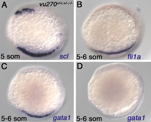
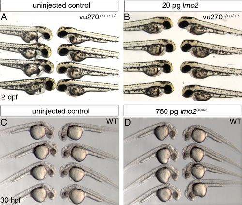
lmo2 mutant zebrafish embryos. (A and B) Heads of normal (A) and lmo2 mutant (B) live embryos at 2 dpf. Arrows point at the optic fissure. (C and D) 30 h post-fertilization (hpf) normal (C) and lmo2 mutant (D) embryos labeled for RBC marker gata1. (E and E′) Chromatograms showing normal lmo2 (E) and mutant lmo2vu270 (E′) coding sequence at the mutation site. (F) Schematic presentation of Lmo2 protein and the site of predicted truncation in Lmo2 encoded by vu270 allele (arrow). (G) Quantification of rescue of lmo2 mutant phenotypes by injection of RNA encoding normal Lmo2 or Lmo2C94X. Numbers are summary of at least two independent experiments. (***p<0.0001). |
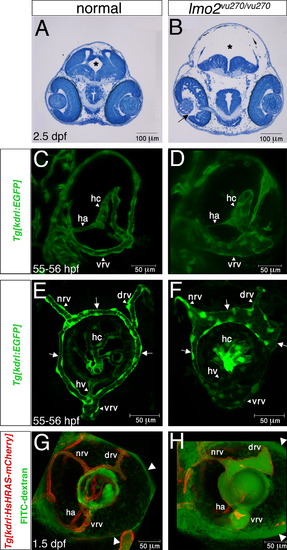
Edema and ocular vessel phenotypes in lmo2 mutant embryos. (A and B) Transverse sections of heads from normal (A) and lmo2 mutant (B) embryos at 2.5 dpf. In the mutant note enlarged ventricle and white spaces within head and eye (arrow) tissues suggesting severe edema. Asterisks mark brain ventricle. (C and D) Three-dimensional shadow rendering confocal images of 55–56 hpf Tg(kdrl:EGFP) normal (C) and lmo2 mutant (D) eyes in frontal view. (E and F) Three-dimensional maximum projection confocal images of 55–56 hpf Tg(kdrl:EGFP) normal (E) and lmo2 mutant (F) eyes in lateral view. Arrows point at different regions of the superficial ring vessel. (G and H) FITC-dextran microangiography of Tg(kdrl:HsHRAS-mCherry) normal (G) and lmo2 mutant (H) eyes in live embryos at 1.5 dpf, lateral view. Note accumulation of dye around the mutant eye (arrowheads). drv, dorsal radial vessel; ha, hyaloid artery; hc, hyaloid capillaries; hv, hyaloid vein; nrv, nasal radial vessel; vrv, ventral radial vessel. In (E–H) anterior is to the left. |

lmo2 is expressed in the developing head vasculature. (A and B) Whole-mount in situ hybridization showing lmo2 expression in wild-type embryos. Arrowheads and arrows point at the ROC and MOC respectively. Asterisks mark the location of eyes. (C–H) 2 μm single confocal sections of combined in situ hybridization for lmo2 expression (red) and antibody labeling for EGFP (blue) in endothelial precursors in Tg(kdrl:EGFP) embryos. lmo2 is expressed in most ROC (C–E) and in some (arrow) MOC (F–H) endothelial precursors. In (F–H) the posterior and medial borders of the eye are outlined. All images are of 15–16 somite-stage embryos. (A) Lateral view and (B-H) dorsal view, anterior to the left. EXPRESSION / LABELING:
|

Optic fissure closure is interrupted at 36 hpf in lmo2 mutant embryos. (A–D) Transmitted light images of eyes of normal (A and C) and lmo2 mutant (B and D) embryos at 30 hpf (A and B) and 36 hpf (C and D). Arrows point at the margins of the optic fissure. All panels are lateral views, anterior to the left. PHENOTYPE:
|
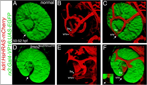
Dilated vessels are located in the optic fissure. (A–F) Three-dimensional shadow rendering of confocal z-stacks of eyes (green) and ocular vessels (red) in 2 dpf (rx3:Gal4-VP16);(UAS:EGFP);(kdrl:HsHRAS-mCherry) transgenic normal (A–C) and lmo2vu270/vu270 (D–F) embryos. In the merged panels (C and F) most of the hyaloid vessels cannot be seen. (E and F) Growth of superficial ring vessel is delayed in the mutant (compare to B and C). Arrows point at the optic fissure. Inset in F is a higher magnification of the optic fissure area with the arrow pointing at the dilated vessel in the fissure. Vessel abbreviations are as in Fig. 2. All panels are dorsolateral view, anterior to the left. |

Ocular vessel dilation is flow dependent and interferes with optic fissure closure. (A–D) Single plane confocal images combined with transmitted light image of eyes of normal Tg(kdrl:EGFP) embryos (A and C) and lmo2vu270/vu270; Tg(kdrl:EGFP) embryos (B,D), uninjected (A and B) or injected with MO1-tnnt2a (C and D). Arrows point at GFP-positive endothelial cells (green) in close contact with optic fissure margins. (A′–D′) Images of the same embryos as in A–D. Arrows point at optic fissure margins. Embryos were treated with PTU to block pigmentation. All panels are lateral view, anterior to the left. |
|
|

|
Reprinted from Developmental Biology, 369(2), Weiss, O., Kaufman, R., Michaeli, N., and Inbal, A., Abnormal vasculature interferes with optic fissure closure in lmo2 mutant zebrafish embryos, 191-198, Copyright (2012) with permission from Elsevier. Full text @ Dev. Biol.