
- Title
-
A zebrafish Notum homolog specifically blocks the Wnt/beta-catenin signaling pathway
- Authors
- Flowers, G.P., Topczewska, J.M., and Topczewski, J.
- Source
- Full text @ Development
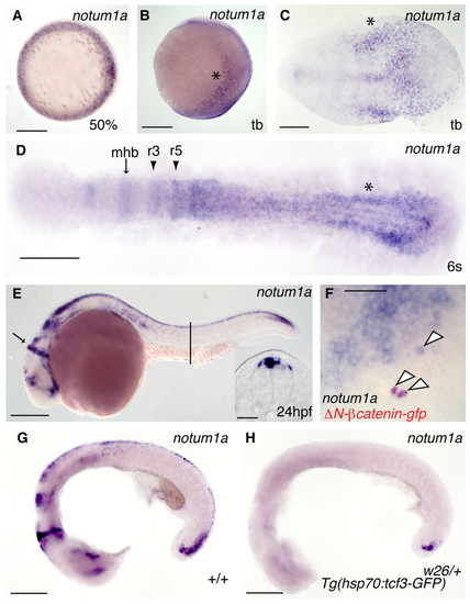
Expression of zebrafish notum1a. (A-E) Expression of zebrafish notum1a. (A) At 50% epiboly, notum1a is expressed around the entire blastoderm margin. (B) A lateral view of notum1a expression at tail bud reveals expression in the lateral neural plate (asterisk). (C) A flat-mount of the tail bud displays notum1a expression in the posterior neural plate, particularly at the lateral edges (asterisk) and midline. (D) At six somites, as neurulation proceeds, lateral neural plate notum1a expression becomes more medially located. More anteriorly, notum1a begins to appear in the midbrain-hindbrain boundary (mhb, arrow) and rhombomeres 3 and 5 (r3, r5, arrowheads). (E) At 24 hpf, notum1a is expressed in the mhb (arrow) and in the dorsal neural tube, as revealed in a section through the trunk at the level of the yolk extension (line, inset). Scale bar: 50 μm. (F,G) Wnt/β-catenin signaling is needed for notum1a expression. (F) Mosaically expressed activated β-catenin (red) colocalizes with ectopic notum1a expression (arrowheads) at 80% epiboly. Scale bar: 50 μm. (G,H) Although normally expressed in heat-shocked non-transgenic embryos at 22 hpf (G), notum1a is absent in heat-shocked Tg(hsp70:tcf3-gfp)w26/+ embryos (H) except in small domain within the tail bud 2 hours post-heat shock. Scale bars in A-D,G,H: 200 μm. EXPRESSION / LABELING:
|

Notum 1a is an inhibitor of Wnt signaling. (A) Overexpression of notum1a produces phenotypes resembling and synergistic with dkk1 overexpression. (Left) Representative embryos following injection of increasing amounts of notum1a. Phenotypic classes are marked by colored squares. (Right) Injection of a quantity of notum1a mRNA that does not produce overt phenotypes enhances the severity of phenotypes seen in embryos injected with dkk1. (B,C) tbx6, which is expressed around the margin at 60% in wild-type embryos (B) is eliminated following notum1a overexpression (C). (D,E) gsc expressed on the presumptive dorsal side of the embryo in wild-type shield stage embryos (D), is expanded following notum1a injection (E). (F) Notum 1a interacts with the Wnt/β-catenin pathway. Injection of wnt8 or wnt1 mRNA or dkk1 MO produces embryos lacking eyes (lightest blue) or with smaller eyes (light blue) than wild-type embryos (dark blue) at 24 hpf. Co-injection of notum1a suppressed wnt8 and wnt1 mRNA, and dkk1 MO-induced eye reduction. MO-mediated depletion of notum1a with a splice-site-directed MO enhanced eye reduction induced by wnt1 mRNA; this enhancement was rescued by injection of notum1a mRNA. (G-J) Stimulation of Wnt signaling by LiCl partially restored wild-type forebrain rx3 expression following notum1a overexpression. (G) Flat-mounted control, non-heat shocked Tg(hs:notum1a) embryo at eight somites with in situ hybridization against rx3 (labeling forebrain), pax2a (midbrain-hindbrain boundary and pronephric mesodern), egr2b (krox20) (rhombomeres three and five) and myod1 (adaxial cells and somites) (H) Tg(hs:notum1a) embryos heat shocked at 50% epiboly display expanded rx3 domain. (I) Eight minutes of LiCl treatment at 75% epiboly eliminated rx3 expression in non-heat-shocked embryos. (J) Heat-shock at 50% epiboly before the same LiCl treatment at 75% epiboly partially restores rx3 expression. PHENOTYPE:
|
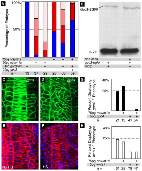
Notum 1a interacts with specific glypicans. (A) Inhibition of posterior body in notum1a-overexpressing embryos is enhanced by depletion of gpc3. Although co-injection of gpc3 MO increases the number of embryos with severe inhibition of posterior body (P=0.003), co-injection of gpc3 mRNA reduces the number of embryos displaying any posterior body impairment compared with notum1a-injected (P=0.05) and notum1a mRNA- and gpc3 MO-injected (P=0.002) embryos. The phenotypic classes are the same as depicted in Fig. 2A. (B) Notum 1a overexpression produces a faster migrating form of Gpc3-EGFP. A western blot of lysates from mGFP/gpc3-EGFP-injected and mGFP/gpc3-EGFP/notum1a-injected embryos probed with an anti-GFP antibody reveals that the gpc3-EGFP band migrates faster in lysates from notum1a-injected embryos. (C-F) Notum 1a overexpression does not mimic the Gpc4 loss-of-function phenotype. (C,D) Confocal images of the notochords of gpc4fr6/+ (C) and gpc4fr6/fr6 (D) Tg(Bactin:HRAS-EGFP)vu119 embryos at five somites. Although the cells of the notochords of gpc4fr6/+ embryos display normal mediolateral elongation and orientation, the cells are disorganized in the homozygous mutants. (E,F). Confocal images of three somite embryos injected with mCherry-HsHRAS RNA non-heat shocked and Tg(hs:notum1a) embryos heat shocked at 50% epiboly. Both control (E) and notum1a-overexpressing (F) embryos display normal cell elongation and orientation. (G) notum1a does not interact with Gpc4 or Wnt11. An incross of gpc4fr6/+ results in a Mendelian ratio of embryos displaying a mutant phenotype at 24 hpf. Injection of gpc4 mRNA efficiently rescues this phenotype. Co-injection of notum1a with gpc4 mRNA does not impair the rescue of the mutant phenotype (P=0.565). (H) Similarly, wnt11tz216/+ fish were incrossed to produce a Mendelian ratio of mutant embryos displaying some degree of cyclopia. This phenotype can be suppressed by injection of wnt11 mRNA, and co-injection of notum1a did not inhibit this effect (P=0.95). PHENOTYPE:
|

Notum 1a is required for proper neural tube patterning. (A,B) Compared with uninjected controls at 24 hpf (A), notum1a expression is expanded in the neural tubes of Notum 1a-depleted embryos (P=7.8 × 10⁻⁹) (B). (C,D) Similarly, compared with uninjected embryos at 24 hpf (C), msxc is expanded in notum1a MO-injected embryos (P=2.0 × 10⁻⁴) (D). (E,F) msxc expression is lost in the dorsal neural tube of heat-shocked Tg(hs:notum1a) embryos at 24 hpf (F). Note the normal expression of ntl in the notochord. (G-J) Loss of notum1a leads to ectopic dkk1 expression in the neural tube. Although dkk1 is not observed in the neural tube of uninjected embryos at 24 hpf (G), it was faintly (I) or intensely (H) expressed within the neural tube in the majority of notum1a MO-injected embryos (J). EXPRESSION / LABELING:
|
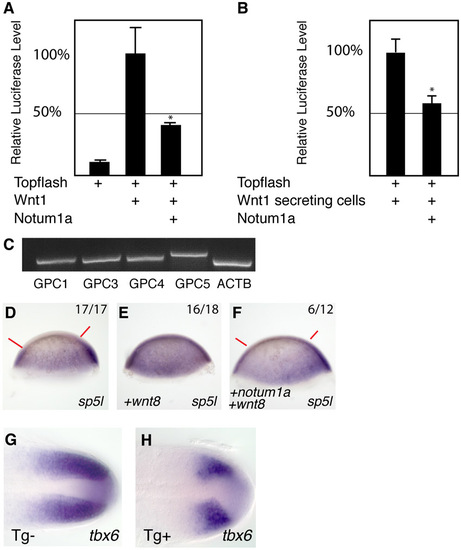
Notum 1a inhibits Wnt/β-catenin signaling. (A-C) Notum 1a inhibits Wnt signaling in cultured cells. (A) Expression of notum1a in HEK 293 cells results in a significant, dose-dependent reduction in the relative activation of the TOPFLASH reporter to zebrafish Wnt1 (P=0.019). Co-transfection of gpc3 with notum1a potentiates the notum1a-mediated inhibition of TOPFLASH activity (P=0.013). (B) Notum1a inhibits reception of Wnt signal. Cells were transfected with wnt1 and, 4 hours post-transfection, plated with cells transfected with either topflash alone or topflash and notum1a. The reporter response of notum1a-transfected cells was significantly reduced (P=0.00002). Bars indicate standard deviations. (C) RT-PCR reveals expression of GPC1, GPC3, GPC4 and GPC5 in HEK293 cells. ACTB serves as a positive control. (D-F) Notum 1a inhibits Wnt-induced expansion of the sp5l domain. (D) sp5l at shield stage is not expressed in the animal pole of the embryo (between red bars) in wild-type embryos. (E) In wnt8-injected embryos, sp5l is ubiquitously expressed. (F) Injection of notum1a suppresses ubiquitous sp5l expression in wnt8-injected embryos (P=0.03). (G,H) Induction of notum1a during somitogenesis reduces tbx6 expression domain. (G) Heat-shocked non-transgenic embryos display normal domain of tbx6 expression at 16 somites. (H) notum1a expression at 16 somites inhibits tbx6 expression in the majority of heat shocked Tg(hs:notum1a) embryos. EXPRESSION / LABELING:
|
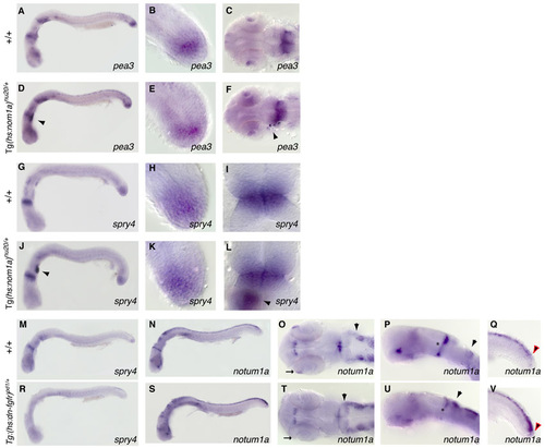
Notum 1a is not part of a feedback loop regulating Fgf signaling. (A-N) Overexpression of notum1a in Tg(hs:notum1a) embryos does not alter FGF signaling. (A-C) Expression of pea3 at 22 hpf in non-transgenic embryos heat shocked at 20 hpf in the whole embryo (A), tail (B) and MHB (C). (D-F) Expression of pea3 at 22 hpf in transgenic embryos heat shocked at 20 hpf is not altered. egfp co-staining in the heart indicates the presence of the transgene (arrowhead). (G-I) Expression of spry4 at 22 hpf in non-transgenic embryos heat shocked at 20 hpf in the whole embryo (G), tail (H) and MHB (I). (J-L) Expression of spry4 at 22 hpf in transgenic embryos heat shocked at 20 hpf is not altered. egfp expression is present in J and L (arrowheads). (M) Expression of spry4 at 24 hpf in non-transgenic embryos heat-shocked at 22 hpf. (N-Q) Expression of notum1a at 24 hpf in non-transgenic embryos heat-shocked at 22 hpf. (O) A dorsal view of the head reveals notum1a expression in the eye (arrow) and the anterior extent of notum1a expression in the hindbrain in rhombomere 3. (P) A lateral view of the head shows MHB extending through the entire dorsoventral axis of the brain (asterisk). (Q) In the tail, notum1a expression diminishes posteriorly (red arrow). (R) At 24 hpf, spry4 expression is absent in heat-shocked Tg(hsp70l:dn-fgfr1-EGFP)pd1/+embryos 2 hours post-heat-shock. (S-V) Expression of notum1a at at 24 hpf in n Tg(hsp70l:dn-fgfr1-EGFP)pd1/+embryos heat-shocked at 22 hpf. Although the majority of notum1a expression is intact following heat-shock (S), anterior eye expression is lost (arrow, T) and expression is expanded into the rhombic lip (arrowhead, T, U). The dorsoventral extent of MHB expression is reduced (asterisk, U). The posterior domain in the tail is expanded (V, red arrow). |

Overexpression of notum1a does not impair endoderm formation. (A,B) Non-heat shocked (A) and heat-shocked (B) Tg(hs:notum1a) embryos display similar levels of sox32 expression at 90% epiboly. |

Notum 1a morphant phenotype. (A) Uninjected embryo at 25 somites. (B) notum1a-MO-injected embryo at 25 somites. (C) notum1a expression in an uninjected embryo at 24 hpf. (D) notum1a is globally expanded in notum1a-MO-injected embryos at 24 hpf. PHENOTYPE:
|
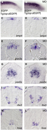
Loss of Notum1a results in expanded Wnt reporter activity, but not bmp4, foxa, pax3a or pax6a expression. (A,B) Relative to that in uninjected controls (A), the domain of egfp expression in the tails of embryos in the Tg(TOP:dEGFP)w26 reporter line is expanded in notum1a MO-injected embryos (B) at 24 hpf. (C-L) Transverse sections of neural tubes at 24 hpf. bmp4 (C,D), pax3a (E,F), pax6a (G,H), lhx3 (I,J) and foxa (K,L) are not affected in notum1a MO-injected embryos. EXPRESSION / LABELING:
|

Unillustrated author statements EXPRESSION / LABELING:
PHENOTYPE:
|