
- Title
-
Zebrafish sox9b is crucial for hepatopancreatic duct development and pancreatic endocrine cell regeneration
- Authors
- Manfroid, I., Ghaye, A., Naye, F., Detry, N., Palm, S., Pan, L., Ma, T.P., Huang, W., Rovira, M., Martial, J.A., Parsons, M.J., Moens, C.B., Voz, M.L., and Peers, B.
- Source
- Full text @ Dev. Biol.

sox9b expression in the HPD system, IPD and IHD throughout zebrafish embryonic development. (A) sox9b transcript is detected in the region of the hepatic and ventral pancreatic primordia (HVP). The yellow arrowhead points to the dorsal pancreas (DP). (B,C) During embryogenesis, sox9b expression delineates a network connecting the liver and pancreas. Yellow dotted lines delimit the pancreas and the liver primordia. (D) Confocal section through sox9b and prox1a expression in the HVP domain at 28 hpf. The brackets highlight the overlap between both expression domains. (E) Confocal section through sox9b and pdx1 adjacent and partially common expression domains. (F) Immunodetection of Prox1 and Pdx1 revealing a partial overlap (bracket). (G) sox9b expression in the ventral pancreas of Tg(ptf1a:eGFP) embryos. (H) Confocal projection of sox9b and the hepatocyte marker cp expression in the hepatic bud at 52 hpf. (I, J) Comparison at 3 dpf of sox9b expression in the liver labelled by cp (I) with the pattern of the IHD labelled with the ductal marker 2F11 and Prox1 (the IHDs appear yellow) (J). sox9b expression is restricted to cp-cells (I, arrows) and displays a highly similar pattern to 2F11+ cholangiocytes (J). (K) At this stage, in the pancreatic acinar tissue labelled with ptf1a, sox9b is restricted to IPD (inset) and EPD cells. (L) Detection of SOX9 protein in the Notch-responsive cells (PNCs) along the IPD of Tg(Tp1:hmgb1-mCherry) transgenic larvae at 5 dpf. (M) sox9b mRNA detection followed by immunolabelling with the ductal maker 2F11 demonstrating sox9b expression in the IHD and in the HPD system, notably in the gall bladder (GB, arrowhead). The dotted lines delineate the border of the liver and pancreas with the HPD. Note that the IPD could not be detected by 2F11 immunolabelling following in situ hybridisation. EHD, extrahepatic duct; EI, endocrine islet; EPD, extrapancreatic duct; GB, gall bladder; HB, hepatic bud; HVP, hepatic and ventral pancreatic domain; IHD, intrahepatic ducts; IPD, intrapancreatic ducts; OV, otic vesicle; VP: ventral pancreas. Scale bar=40 μm except H, I and J=20 μm. EXPRESSION / LABELING:
|

Affected morphogenesis of the IPD, IHD and HPD system in sox9b mutants. (A, B) Acinar (try) and hepatocyte (tfa) differentation as well as the global morphology of the larvae are similar in wild type larvae (A) and in sox9b mutants (B) at 84 hpf. (C, D) Three dimensional rendering of the liver and pancreas at 84 hpf, labelled by Prox1, and of the entire ductal network highlighted by 2F11. The insets represent the 2F11 staining in the liver. White arrows in D and inset point at the disrupted connections between cholangiocytes in sox9b mutants. (E, F) Close-up of the HPD system connecting the pancreas and the liver to the intestine. Ectopic Prox1+ cells are detected throughout the HPD system (red arrow). (E2, F2) Less interconnecting ducts are detected in the IHD sox9b mutants. (E3, F3) 3-D rendering of the pancreas showing weaker 2F11 labelling in the IPD of sox9b mutant (F3) compared to wild type (E3). (G, H). 3-D rendering at 7 dpf of ductal 2F11 labelling in the liver (IHD) in wild type (G) and sox9b mutants (H). (G2, H2) Higher magnification showing thicker connections between IHD cells (asterisk). (G3, H3) 3-D rendering at 7 dpf of ductal 2F11 labelling in the pancreas (IPD) in wild type (G2) and sox9b mutants (H2). GB, gall bladder; HPD, hepatopancreatic ductal system; IHD, intrahepatic ducts; IPD, intrapancreatic ducts. Scale bar=20 μm. EXPRESSION / LABELING:
PHENOTYPE:
|
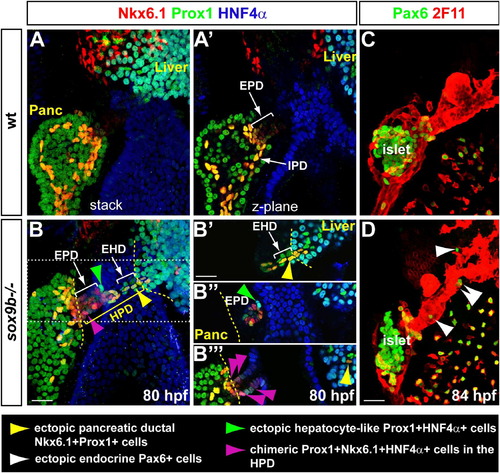
Misdifferentiation of the HPD system in sox9b mutants. (A, B) 3D-rendering (stack) at 80 hpf of the liver, pancreas and HPD system immunolabelled with Prox1, HNF4α and the pancreatic ductal marker Nkx6.1. (A2, B2–B32) Z-planes through the same larvae as in A and B. Prox1 with HNF4α label hepatocytes while Prox1 together with Nkx6.1 strongly labels the IPD. In sox9b mutants (B and B2–B32), the HPD system becomes labelled with the three markers and display different ectopic cell types (see the colour code at the bottom). Dotted lines delimitate organs and ducts boundaries. (C, D) 3-D rendering of the HPD system at 84 hpf labelled with the ductal marker 2F11 and the endocrine marker Pax6. Ectopic endocrine cells along the HPD and in the liver are indicated by white arrowheads. EHD, extrahepatic duct; EPD, extrapancreatic duct; HPD, hepatopancreatic ductal system; IPD, intrapancreatic ducts. Scale bar=20 μm. EXPRESSION / LABELING:
PHENOTYPE:
|

sox9b early hepatopancreatic expression is activated by FGF and BMP and is maintained in the ducts by FGF and Notch. (A, B) sox9b early expression (30 hpf) in the hepatic and ventral pancreatic primordia (HVP) is not activated in compound fgf10; fgf24 mutants (C, D) Similarly, sox9b is not induced in bmp2a morphants. Note that sox9b in the dorsal pancreatic bud (DP) is not affected. |

Concomitant maintenance of ductal sox9b expression with repression of endocrine differentiation by FGF and Notch signalling. (A, B) Expression at 3 dpf of sox9b with the endocrine marker neurod in wild type embryos treated from 54 to 72 hpf with DMSO (A) or with the FGF inhibitor SU5402 (B). Confocal planes containing the EPD are shown. (A2, B2) sox9b expression only is shown. (C, D) Ectopic Nkx6.1+ and endocrine Pax6 cells are found in the HPD of fgf10 mutants, as in sox9b mutants. Yellow dotted lines demarcate organ boundaries. (E–F2) 3-D rendering of the pancreas showing sox9b and neurod expression at 4 dpf at the level of the IPD in wild type embryos treated from 72 to 96 hpf with DMSO (E, E2) or with the Notch inhibitor DAPT (F, F2). (E2, F2) sox9b expression alone is shown. (G–G3) Detection of SOX9 protein in the Notch-responsive cells (PNCs) revealed with anti-mCherry in Tg(Tp1:hmgb1-mCherry) transgenic larvae at 5 dpf. (H) Quantitative RT-PCR analyses of sox9b and amylase expression in centroacinar and acinar cells isolated from the pancreas of adult Tg(Tp1:hmgb1-eGFP) (Tp1+) and Tg(ptf1a:eGFP) (ptf1a+) fish, respectively. EPD, extrapancreatic duct; HPD, hepatopancreatic ductal system; IPD, intrapancreatic duct. Scale bar=20 μm. EXPRESSION / LABELING:
PHENOTYPE:
|

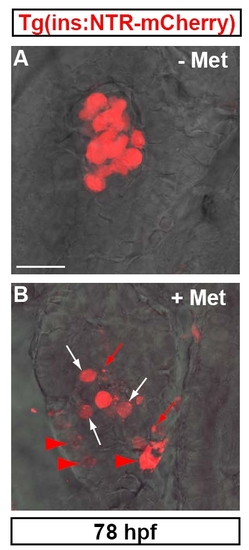
sox9b is required for late endocrine cell formation in the IPD and for islet beta cell recovery upon ablation in larvae. (A–D) Immunodetection at 6 dpf of endocrine cell differentiation (Pax6) along the IPD labelled with 2F11 in wild type (A) and sox9b mutant larvae (B). Larvae were treated with DMSO (A, B) or DAPT (C, D) from 3 to 6 dpf. Secondary isolated endocrine cells are detected (small arrowheads) in wild type and sox9b mutant control larvae. The dotted line demarcates the intestine from the pancreatic region. (C, D) The massive increase in secondary islets resulting from DAPT treatment of wild type larvae (large arrowheads in C) is not observed in sox9b mutants which still display isolated endocrine cells along the IPD (small arrowheads in D). (E–H) Beta cell recovery in Tg(ins:nfsB-mCherry); sox9b larvae upon Metronidazole (Met) mediated ablation. Pancreatic ducts were labelled with 2F11 and beta cells were revealed by Insulin (Ins) immunodetection as mCherry fluorescence was undetectable after the process of immunofluorescence. (E, F) 6.5 dpf Tg(ins:nfsB-mCherry) wild type (E) and sox9b mutant larvae (F) 3 days (80 h) after DMSO treatment from 56 to 80 hpf. (G, H). Tg(ins:nfsB-mCherry) wild type (G) and sox9b mutant (H) larvae 3 days after Met exposure. Scale bar=20 μm. |

EXPRESSION / LABELING:
|
|

Unillustrated author statements |
Reprinted from Developmental Biology, 366(2), Manfroid, I., Ghaye, A., Naye, F., Detry, N., Palm, S., Pan, L., Ma, T.P., Huang, W., Rovira, M., Martial, J.A., Parsons, M.J., Moens, C.B., Voz, M.L., and Peers, B., Zebrafish sox9b is crucial for hepatopancreatic duct development and pancreatic endocrine cell regeneration, 268-278, Copyright (2012) with permission from Elsevier. Full text @ Dev. Biol.