
- Title
-
Nuclear progesterone receptors are up-regulated by estrogens in neurons and radial glial progenitors in the brain of zebrafish
- Authors
- Diotel, N., Servili, A., Gueguen, M.M., Mironov, S., Pellegrini, E., Vaillant, C., Zhu, Y., Kah, O., and Anglade, I.
- Source
- Full text @ PLoS One
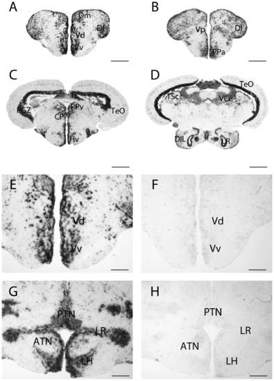
Expression of pgr transcripts in the forebrain of adult zebrafish. A, B, C and D: pgr transcripts detection with antisense probes in the telencephalon (A), the preoptic area (B), in the ventromedial hypothalamus (C) and the nucleus recessus posterioris (D). E, F, G and H: In situ hybridization with antisense probes (E and G) and sense probes (F and H) on parallel sections. The sense probe does not generate any signal. ATN: anterior tuberal nucleus; CP: central posterior thalamic nucleus; DIL: diffuse nucleus of the inferior lobe; Dl: Lateral zone of the dorsal telencephalic area; Dm: medial zone of the dorsal telencephalic area; Hv: ventral zone of the periventricular hypothalamus; LH: lateral hypothalamic nucleus; LR: Lateral recess; PTN: posterior tuberal nucleus; Vv: ventral nucleus of the ventral telencephalic area; Vd: dorsal nucleus of the ventral telencephalic area; PGZ: periventricular gray zone of the optic tectum; PPv: periventricular pretectal nucleus; TeO: Optic tectum; TL: torus longitudinalis; TSc: torus semicircularis; VCe: valvula cerebella. Scale bars: 50 μm (G and H); 100 μm (E and F); 200 μm (A and B); 400 μm (C and D). EXPRESSION / LABELING:
|
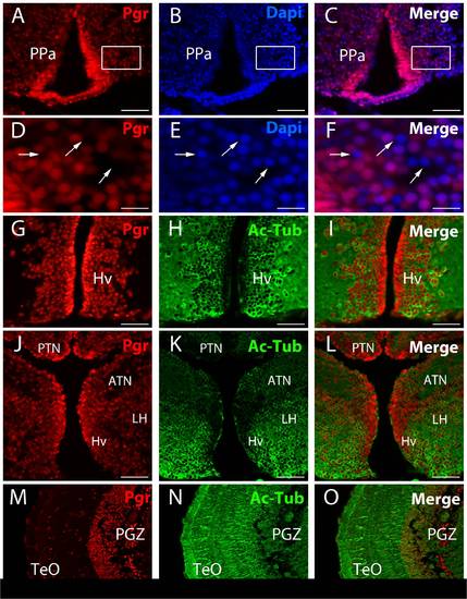
Immunolocalisation of Pgr expressing neurons in adult zebrafish forebrain. A to F: Pgr immunohistochemistry (red) with DAPI nuclei staining (blue) showing that not all nuclei express Pgr (arrows). D, E and F correspond to high magnifications of the area framed in A, B and C. Arrows point to nuclei that do not express Pgr. G to O: Pgr (red) and acetylated-tubulin (green) staining showing that numerous Pgr positive cells correspond to acetylated-tubulin positive neurons. Note that cells lining the ventricle are strongly positive for the Pgr antibodies, but are not stained by acetylated-tubulin. ATN: anterior tuberal nucleus; Hv: ventral zone of the periventricular hypothalamus; LH: lateral hypothalamic nucleus; PGZ: periventricular gray zone of the optic tectum; PPa: anterior part of the preoptic area; TeO: Optic tectum. Scale bars: 15 μm (D, E and F), 50 μm (A, B, C, G, H, I, J, K and L), 100 μm (M, N and O) EXPRESSION / LABELING:
|

Radial glial cells strongly express Pgr. A to O: Pgr immunohistochemistry (red) on tg(cyp19a1b-GFP) transgenic fish showing the strong expression of Pgr in cyp19a1b-GFP positive radial glial cells (green). The cyp19a1b-GFP radial glial cells (green) that line the ventricular surface exhibit a stronger Pgr staining, notably in the subpallial (A to F) and pallial regions (G to I). This is also true in more posterior regions such as the posterior tuberculum (J to K) or around the posterior recess (NPR) of the caudal hypothalamus (M to O). Vv: ventral nucleus of the ventral telencephalic area; Vd: dorsal nucleus of the ventral telencephalic area; Dm: medial zone of the dorsal telencephalic area; TPp: periventricular nucleus of the posterior tuberculum; NPR: nucleus of the posterior recess of the hypothalamus Scale bars: 30 μm (D, E and F); 75 μm (J, K and L); 150 μm (A, B, C, G, H, I, M, N and O). EXPRESSION / LABELING:
|
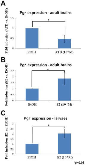
Nuclear progestin receptor is up-regulated by estrogen in both the developing and adult brain. Fold induction of pgr expression after treatment of adult zebrafish with an aromatase inhibitor (A, 10-6 M of ATD) or estradiol (B, 10-7 M of 17β-estradiol), and larvae with E2 (C, 10-8 M of 17β-estradiol). In A, the aromatase inhibitor (ATD) leads to a significant decrease of pgr expression in individual brains of adult zebrafish (n = 4). In B, the estrogenic treatment leads to a significant increase of pgr expression in pools of 5 brains of adult zebrafish (n = 3). In C, the estrogenic treatment leads to a significant increase of pgr expression in pool of 20 larvae (n = 2). Asterisk (*) indicates significant differences (p<0.05, Student′s t test). The graphs present the mean value +/- the standard deviation. EXPRESSION / LABELING:
|

Nuclear progestin receptors are up-regulated by estradiol. A to P: Pgr immunohistochemistry (red) on tg(cyp19a1b-GFP) transgenic larvae (green) with DAPI counterstaining (blue) in the anterior (PPa) and posterior (PPp) preoptic area in two different control larvae (A–D and I–L) and two E2-treated larvae (E–H and N–P). A–D: Control larva treated with EtOH: Although the Pgr staining is widely distributed, not all cells are positive as shown by DAPI counterstaining. Only a few cyp19a1b-GFP expressing radial glial cells are visible and some of them express Pgr (D). E–H: Larva treated with 10-8 M of 17β-estradiol: At exactly the same level than in control (A–D), one can see that a majority of cells express Pgr as shown by the DAPI counterstaining. Furthermore, Pgr staining intensity is stronger than in controls. The estrogenic treatment also leads to an increase in the number of cyp19a1b-GFP positive radial glial cells expressing Pgr (H). Pictures in E and F were taken with the same exposure time than A and B, respectively. I–L: Control larva treated with EtOH shown at higher magnification in the posterior preoptic area. The Pgr staining is clearly detectable in some cells as indicated by the DAPI counterstaining. As shown by the merge picture in L, the cyp19a1b-GFP radial glial cells do not express Pgr. M–P: Larva treated with 10-8 M of 17β-estradiol (E2) in the exactly the same region than in I–L. Pgr is expressed in a large majority of cells and the staining intensity is clearly much stronger than in control larvae. Furthermore, all cyp19a1b-GFP positive radial glial cells also express Pgr. Pictures in M and N were taken with the same exposure time than I and J, respectively. Scale bars: 60 μm (A, B, C, D, E, F, G and H); 30 μm (I, J, K, L, M, N and O). EXPRESSION / LABELING:
|