
- Title
-
Identification of RNA binding motif proteins essential for cardiovascular development
- Authors
- Maragh, S., Miller, R.A., Bessling, S.L., McGaughey, D.M., Wessels, M.W., de Graaf, B., Stone, E.A., Bertoli-Avella, A.M., Gearhart, J.D., Fisher, S., and McCallion, A.S.
- Source
- Full text @ BMC Dev. Biol.
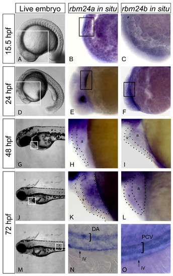
rbm24a and rbm24b display cardiovascular expression during embryogenesis. Expression of rbm24 transcripts was evaluated in uninjected embryos fixed at 15.5, 24, 48 and 72 hpf. Live embryos 15.5 hpf with area of interest for rbm24 in situ boxed in white (A). Lateral heart views of rbm24a and rbm24b in situs on uninjected 15.5 hpf embryos showed expression linearly organized myocardial precursor cells for rbm24a (black box, B) but not rbm24b (C). Live embryos 24 hpf with area of interest for rbm24 in situ boxed in white (D). Lateral heart views of rbm24a and rbm24b in situs showed expression in the developing heart tube at 24 hpf (black box, E, F) with lens expression for rbm24a alone. 48 hpf live embryo showing the heart boxed in white (G). Lateral zoom of the heart showed rbm24a and rbm24b were expressed in the ventricle (v) and atrium (a) of the looped heart at 48 hpf (H, I). Live 72 hpf embryos with the heart boxed in white (J). Expression of both rbm24 transcripts was detected in the heart at 72 hpf (K, L). Live image of a 72 hpf embryo with the area of interest for vascular expression boxed in white (M). Expression of both rbm24 transcripts was detected in the trunk vasculature with differing expression patterns. rbm24a shows arterial expression in the DA (N) while rbm24b shows venous expression in the PCV (O) with both being expressed in the IV. DA, dorsal aorta; PCV, posterior caudal vein; IV, intestinal vasculature. EXPRESSION / LABELING:
|
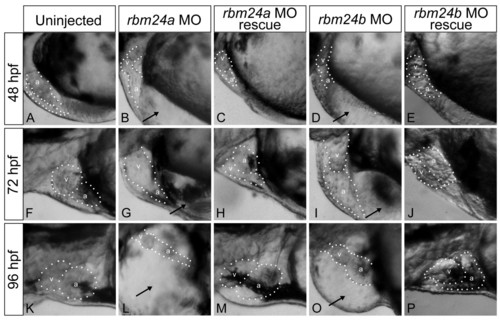
rbm24a and rbm24b are required for normal cardiac development. Translation blocking morpholinos complementary to rbm24a (5 ng) or rbm24b (8 ng) were injected into 1-2 cell stage zebrafish embryos and the resulting phenotypes were evaluated compared to uninjected controls at 48 hpf (A, B, D), 72 hpf (F,G,I), and 96 hpf (K, L, O). Lateral heart views are shown with a dotted outline around the heart chambers. Both morphant embryo conditions exhibited cardiac looping defects and edema at all stages. Heart chambers are shown with a dotted outline with chambers denoted: v, ventricle; a, atrium; black arrows, cardiac edema. Phenotype rescue was achieved for each rbm24 via co-injection of each respective full length capped poly-A RNA transcript (rbm24a 800 pg, rbm24b 50 pg) along with the respective complementary translation blocking morpholino into 1-2 cell stage embryos where 800 pg rbm24a (C, H, M) or 50 pg rbm24b (E, J, P) achieved rescue. Rescued embryos posses looped hearts absent of edema. |

Depletion of rbm24a and rbm24b compromise cardiac myocardial development. The expression patterns of cardiac markers were analyzed in rbm24 morphants and uninjected controls at 72 hpf. Ventral and lateral images of the heart are shown with a dotted line marking the boundary between heart chambers. By myl7 expression of the entire heart, uninjected controls possess looped hearts with defined ventricle and atrium chambers (A, B) in contrast to unlooped hearts for both rbm24 morphants. rbm24a morphants displayed linear presumptive ventricle and an incompletely formed presumptive atrium (C, D). rbm24b morphants displayed a linear presumptive atria and ventricle (E, F). myh6 marking the atrium displayed a looped atrium in controls (G, H) with linear atrium in rbm24a (I, J), and rbm24b morphants (K, L). vmhc expression demarks looped ventricles in controls (M, N). rbm24a morphants displayed little expression bound to an unlooped heart tube where expression extends to the presumptive atrium (O, P). Almost no vmhc expression is detectable in rbm24b morphants (Q, R). v, ventricle region; double arrow, ventricular expression a, atrium region; solid black arrow, atrial expression. |
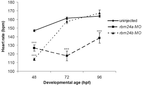
Slow heart rate may contribute to cardiac edema in rbm24 morphants. Heart rates were determined for uninjected control, rbm24a and rbm24b morphant embryos at 48, 72 & 96 hpf and significance was ascribed from a t-test statistic. 50 embryos were examined for each condition at each time point. At all time-points the heart rate of rbm24a morphants was significantly lower than controls where P < 2 × 10⁻⁵. At 48 hpf rbm24b morphants had significantly lower heart rates than controls (P = 1 × 10-32). The heart rates for rbm24b morphants then rose to rates not different from controls by 72 hpf and 96 hpf (P > 0.12) while maintaining the features of the morphant phenotype. Error bars here are standard error. *** P < 0.001. EXPRESSION / LABELING:
|
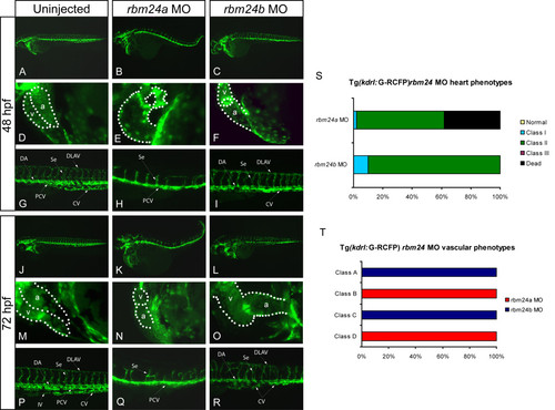
Reduction of rbm24a or rbm24b results in aberrant vasculature and a lack of vascular maintenance. TG(kdrl:G-RCFP) zebrafish line was used to asses the impact of rbm24 depletion on vasculature at 48 hpf and 72 hpf. Lateral views are shown for whole embryo, heart and trunk with a dotted outline indicating heart chamber boundaries. Uninjected controls at all time-points display looped hearts and properly developed vasculature. (A, D, G, J, M, P). 48 hpf rbm24a morphants possess unlooped hearts but display only the PCV and stunted disorganized Se (B, E, H). 48 hpf rbm24b morphants possess unlooped hearts, and only the DA and CV are visible with disorganized Se (C, F, I). By 72 hpf there was increased vascular disorganization and degeneration and a lack of heart looping and in both rbm24 morphants (K, N, L, O, Q, R). Cardiac morphant phenotypes are displayed quantitatively as percentages; Normal, looped beating heart with no cardiac edema; Class I, looped beating heart with cardiac edema. Class II, unlooped beating heart tube with cardiac edema, Class III, beating heart cell mass with cardiac edema; Dead, extreme cell death and degradation of embryo (S). Morphant vascular phenotype classes; Class A, disorganized trunk vasculature with formed DLAV; Class B, disorganized trunk vasculature with truncated Se and no DLAV; Class C, no discernable PCV; Class D, no discernable DA or CV (T). v, ventricle; a, atrium; DA, dorsal aorta; CV, caudal vein; PCV, posterior caudal vein; Se, intersegmental vessels, DLAV, dorsal longitudinal anastomotic vessels; IV, intestinal vasculature. PHENOTYPE:
|
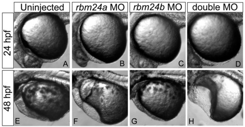
Double rbm24a and rbm24b morphants display more severe cardiac defects. Morpholinos directed to each rbm24 ortholog were injected individually and in tandem into zebrafish embryos and assessed at 24 hpf and 48 hpf compared to uninjected embryo hearts (A and E). Individual morpholino injections were titrated to a low dose for both rbm24a, 2.5 ng (B and F) and rbm24b, 4ng (C and G). Embryos receiving low doses of both rbm24a and rbm24b morpholinos resulted in more severe phenotype than either low dose MO alone, exhibiting severe cardiac defects and increased edema (D and H). |
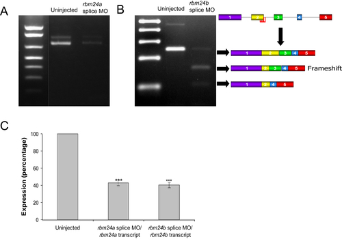
rbm24a and rbm24b splice blocked morphants display cardiac defects. Injection of 7.5 ng of rbm24a splice blocking morpholino results in a substantial reduction of full-length transcript (A). Injection of 9 ng of rbm24b splice blocking morpholino results in aberrant splicing of the transcript. There is a reduction of full-length transcript 250 bp fragment and appearance of trace amounts of the shortened frameshift fragment 164 bp and shortened in-frame transcript 109 bp (B). RT-PCR measurement of transcript levels show both rbm24a (42.80% +/- 3.35, P < 6.5 × 10⁻⁴) and rbm24b (40.27 +/- 3.19, P < 4.4 × 10⁻⁵) morphants have significant reduction of transcript levels compared to uninjected controls (C) Error bars are standard deviation, *** P < 0.001.
|
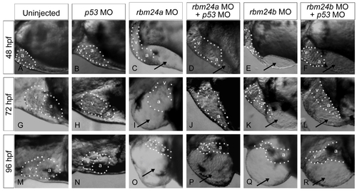
p53 MO co-injection does not alter rbm24a and rbm24 morphant phenotypes. Phenotypes were evaluated for embryos post injection rbm24a MO (5 ng) or rbm24b MO (8 ng) alone or in conjunction with p53 MO (1 ng) phenotypes were compared at 48, 72 & 96 hpf. Lateral heart views are shown with a dotted outline around embryo heart chambers. No cardiac phenotype is detected for p53 MO embryos compared to uninjected controls at any time point (A-B, G, H, M, N). At all time points rbm24a morphants maintain unlooped hearts and display cardiac edema in the presence of p53 MO (C, D, I, J, O, P). Morphant phenotype was also maintained between rbm24b morphants in the presence of p53 MO (E, F, K, L, Q, R). v, ventricle; a, atrium; black arrows, cardiac edema.
|